Veterans Affairs Northern California Health Care System, Martinez, CA, USA.
BMC Med Imaging. 2009 Dec 31;9:20. doi: 10.1186/1471-2342-9-20.
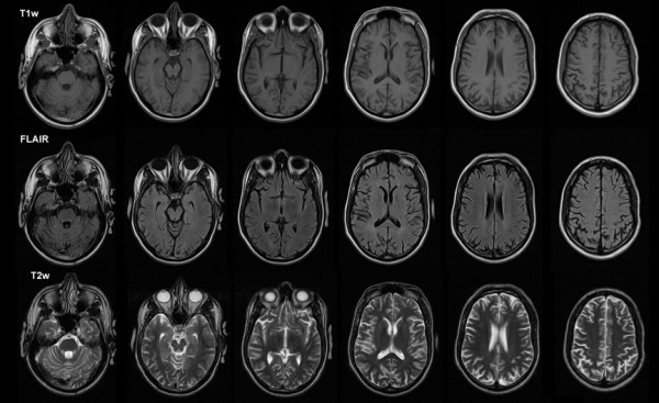
Patients with traumatic brain injury (TBI) often present with significant cognitive deficits without corresponding evidence of cortical damage on neuroradiological examinations. One explanation for this puzzling observation is that the diffuse cortical abnormalities that characterize TBI are difficult to detect with standard imaging procedures. Here we investigated a patient with severe TBI-related cognitive impairments whose scan was interpreted as normal by a board-certified radiologist in order to determine if quantitative neuroimaging could detect cortical abnormalities not evident with standard neuroimaging procedures.
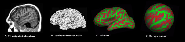
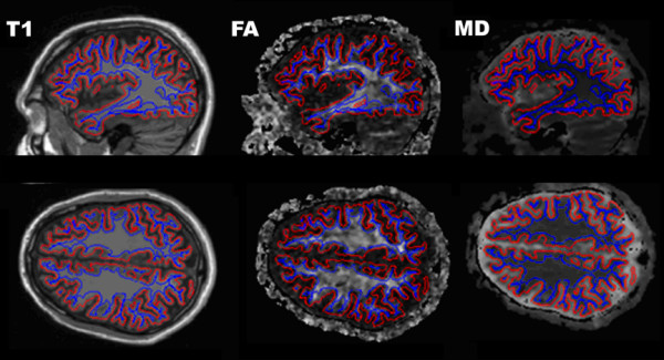
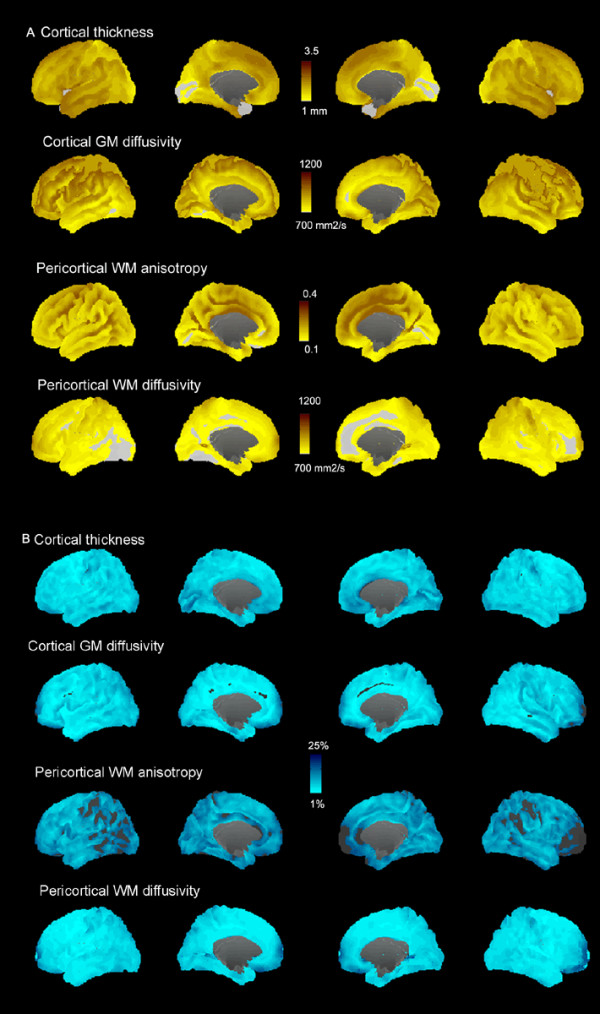
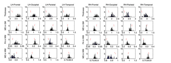
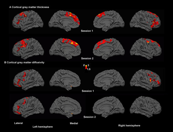
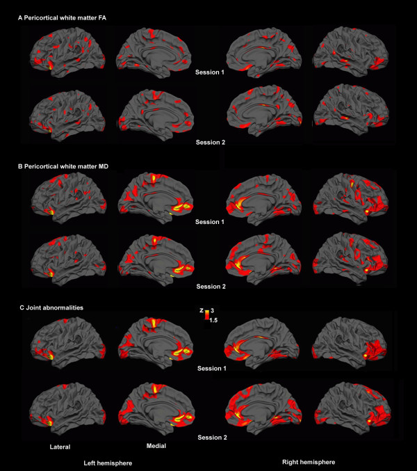
Cortical abnormalities were quantified using multimodal surfaced-based morphometry (MSBM) that statistically combined information from high-resolution structural MRI and diffusion tensor imaging (DTI). Normal values of cortical anatomy and cortical and pericortical DTI properties were quantified in a population of 43 healthy control subjects. Corresponding measures from the patient were obtained in two independent imaging sessions. These data were quantified using both the average values for each lobe and the measurements from each point on the cortical surface. The results were statistically analyzed as z-scores from the mean with a p < 0.05 criterion, corrected for multiple comparisons. False positive rates were verified by comparing the data from each control subject with the data from the remaining control population using identical statistical procedures.
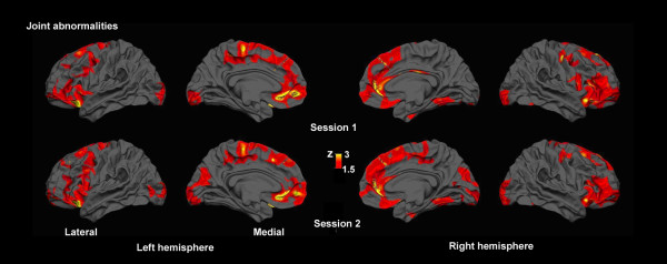
The TBI patient showed significant regional abnormalities in cortical thickness, gray matter diffusivity and pericortical white matter integrity that replicated across imaging sessions. Consistent with the patient's impaired performance on neuropsychological tests of executive function, cortical abnormalities were most pronounced in the frontal lobes.
MSBM is a promising tool for detecting subtle cortical abnormalities with high sensitivity and selectivity. MSBM may be particularly useful in evaluating cortical structure in TBI and other neurological conditions that produce diffuse abnormalities in both cortical structure and tissue properties.
创伤性脑损伤(TBI)患者常表现出明显的认知缺陷,但神经影像学检查无相应的皮质损伤证据。对于这种令人费解的观察结果,一种解释是,TBI 特征性的弥漫性皮质异常难以用标准成像程序检测到。在这里,我们研究了一名严重 TBI 相关认知障碍患者,其扫描结果被一名经过委员会认证的放射科医生解读为正常,以确定定量神经影像学是否可以检测到标准神经影像学程序无法发现的皮质异常。
使用多模态表面形态计量学(MSBM)来量化皮质异常,该方法从高分辨率结构 MRI 和弥散张量成像(DTI)中统计合并信息。在 43 名健康对照者的人群中量化了皮质解剖和皮质及皮质下 DTI 特性的正常值。在两个独立的成像阶段获得了患者的对应测量值。这些数据使用每个脑叶的平均值和皮质表面上的每个点的测量值进行量化。使用均值的 z 分数进行统计分析,并使用 p < 0.05 的标准进行多重比较校正。通过使用相同的统计程序将每个对照受试者的数据与剩余对照人群的数据进行比较,验证了假阳性率。
TBI 患者的皮质厚度、灰质弥散度和皮质下白质完整性存在显著的区域性异常,且在两次成像过程中均有重现。与患者在执行功能神经心理学测试中的表现受损一致,皮质异常在额叶最为明显。
MSBM 是一种有前途的工具,可用于高灵敏度和选择性地检测细微的皮质异常。MSBM 可能特别有助于评估 TBI 和其他引起皮质结构和组织特性弥漫性异常的神经状况的皮质结构。