Shikata Hidenori, McLennan Geoffrey, Hoffman Eric A, Sonka Milan
Iowa Institute for Biomedical Imaging, The University of Iowa, Iowa City, IA 52242, USA.
Int J Biomed Imaging. 2009;2009:636240. doi: 10.1155/2009/636240. Epub 2009 Dec 14.
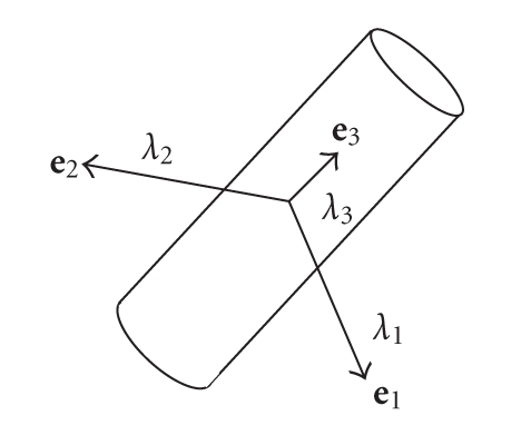
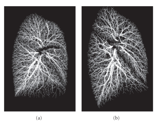
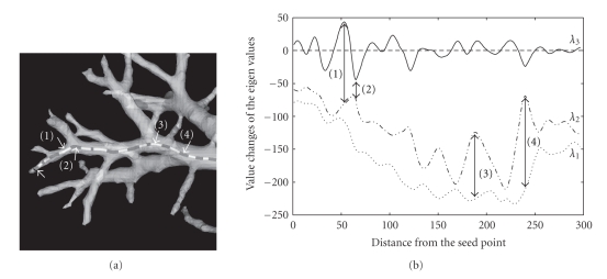
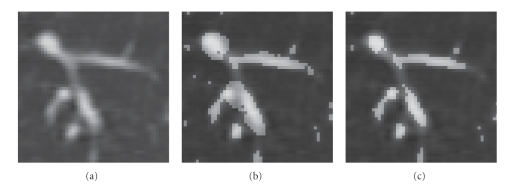
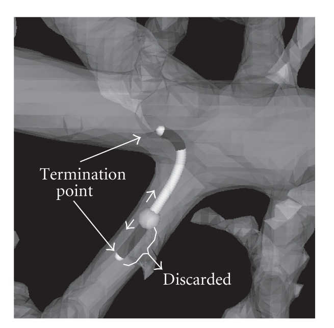
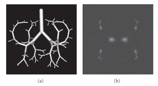
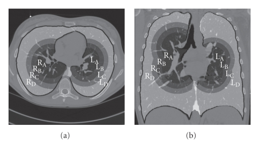
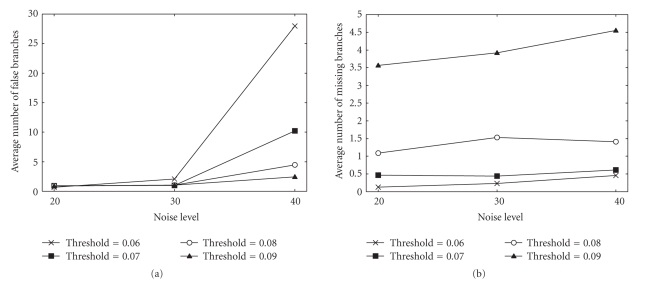
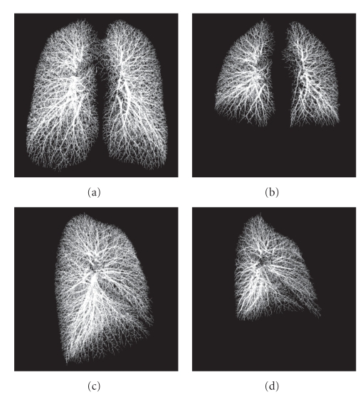
This paper describes an algorithm for extracting pulmonary vascular trees (arteries plus veins) from three-dimensional (3D) thoracic computed tomographic (CT) images. The algorithm integrates tube enhancement filter and traversal approaches which are based on eigenvalues and eigenvectors of a Hessian matrix to extract thin peripheral segments as well as thick vessels close to the lung hilum. The resultant algorithm was applied to a simulation data set and 44 scans from 22 human subjects imaged via multidetector-row CT (MDCT) during breath holds at 85% and 20% of their vital capacity. A quantitative validation was performed with more than 1000 manually identified points selected from inside the vessel segments to assess true positives (TPs) and 1000 points randomly placed outside of the vessels to evaluate false positives (FPs) in each case. On average, for both the high and low volume lung images, 99% of the points was properly marked as vessel and 1% of the points were assessed as FPs. Our hybrid segmentation algorithm provides a highly reliable method of segmenting the combined pulmonary venous and arterial trees which in turn will serve as a critical starting point for further quantitative analysis tasks and aid in our overall goal of establishing a normative atlas of the human lung.
本文描述了一种从三维(3D)胸部计算机断层扫描(CT)图像中提取肺血管树(动脉加静脉)的算法。该算法集成了基于黑塞矩阵特征值和特征向量的管状增强滤波器和遍历方法,以提取细小的外周段以及靠近肺门的粗大血管。所得算法应用于一个模拟数据集以及22名人类受试者在肺活量的85%和20%屏气时通过多排探测器CT(MDCT)成像的44次扫描。通过从血管段内部选择1000多个手动识别的点进行定量验证,以评估真阳性(TPs),并在每种情况下随机在血管外部放置1000个点来评估假阳性(FPs)。平均而言,对于高容量和低容量肺图像,99%的点被正确标记为血管,1%的点被评估为假阳性。我们的混合分割算法提供了一种高度可靠的方法来分割肺静脉和动脉树的组合,这反过来将作为进一步定量分析任务的关键起点,并有助于我们建立人类肺规范图谱的总体目标。