Weinberg Eli J, Mack Peter J, Schoen Frederick J, García-Cardeña Guillermo, Kaazempur Mofrad Mohammad R
Department of Bioengineering, University of California, Berkeley, CA 94720, USA.
Cardiovasc Eng. 2010 Mar;10(1):5-11. doi: 10.1007/s10558-009-9089-9.
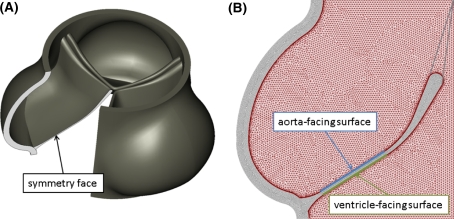
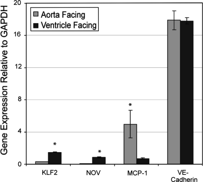
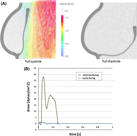
The regulation of valvular endothelial phenotypes by the hemodynamic environments of the human aortic valve is poorly understood. The nodular lesions of calcific aortic stenosis (CAS) develop predominantly beneath the aortic surface of the valve leaflets in the valvular fibrosa layer. However, the mechanisms of this regional localization remain poorly characterized. In this study, we combine numerical simulation with in vitro experimentation to investigate the hypothesis that the previously documented differences between valve endothelial phenotypes are linked to distinct hemodynamic environments characteristic of these individual anatomical locations. A finite-element model of the aortic valve was created, describing the dynamic motion of the valve cusps and blood in the valve throughout the cardiac cycle. A fluid mesh with high resolution on the fluid boundary was used to allow accurate computation of the wall shear stresses. This model was used to compute two distinct shear stress waveforms, one for the ventricular surface and one for the aortic surface. These waveforms were then applied experimentally to cultured human endothelial cells and the expression of several pathophysiological relevant genes was assessed. Compared to endothelial cells subjected to shear stress waveforms representative of the aortic face, the endothelial cells subjected to the ventricular waveform showed significantly increased expression of the "atheroprotective" transcription factor Kruppel-like factor 2 (KLF2) and the matricellular protein Nephroblastoma overexpressed (NOV), and suppressed expression of chemokine Monocyte-chemotactic protein-1 (MCP-1). Our observations suggest that the difference in shear stress waveforms between the two sides of the aortic valve leaflet may contribute to the documented differential side-specific gene expression, and may be relevant for the development and progression of CAS and the potential role of endothelial mechanotransduction in this disease.
人们对人体主动脉瓣血流动力学环境对瓣膜内皮细胞表型的调节了解甚少。钙化性主动脉瓣狭窄(CAS)的结节性病变主要发生在瓣膜小叶主动脉表面下方的瓣膜纤维层中。然而,这种区域定位的机制仍不清楚。在本研究中,我们将数值模拟与体外实验相结合,以研究以下假设:先前记录的瓣膜内皮细胞表型差异与这些个体解剖位置特有的不同血流动力学环境有关。创建了一个主动脉瓣的有限元模型,描述了整个心动周期中瓣膜尖和瓣膜内血液的动态运动。在流体边界上使用具有高分辨率的流体网格,以准确计算壁面剪应力。该模型用于计算两种不同的剪应力波形,一种用于心室表面,另一种用于主动脉表面。然后将这些波形实验性地应用于培养的人内皮细胞,并评估几种病理生理相关基因的表达。与受到代表主动脉面的剪应力波形作用的内皮细胞相比,受到心室波形作用的内皮细胞显示“抗动脉粥样硬化”转录因子Kruppel样因子2(KLF2)和基质细胞蛋白肾母细胞瘤过表达蛋白(NOV)的表达显著增加,而趋化因子单核细胞趋化蛋白-1(MCP-1)的表达受到抑制。我们的观察结果表明,主动脉瓣小叶两侧剪应力波形的差异可能导致已记录的两侧特异性基因表达差异,并且可能与CAS的发生和发展以及内皮机械转导在该疾病中的潜在作用有关。