Beckman Institute for Advanced Science and Technology, University of Illinois, Urbana, IL 61801, USA.
Phys Med Biol. 2010 Feb 21;55(4):1189-201. doi: 10.1088/0031-9155/55/4/019. Epub 2010 Feb 2.
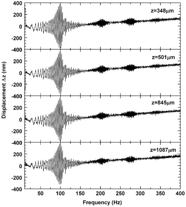
We present a new method for performing dynamic elastography of soft tissue samples. By sensing nanoscale displacements with optical coherence tomography, a chirped, modulated force is applied to acquire the mechanical spectrum of a tissue sample within a few seconds. This modulated force is applied via magnetic nanoparticles, named 'nanotransducers', which are diffused into the tissue, and which contribute negligible inertia to the soft tissue mechanical system. Using this novel system, we observed that excised tissues exhibit mechanical resonance modes which are well described by a linear damped harmonic oscillator. Results are validated by using cylindrical tissue phantoms of agarose in which resonant frequencies (30-400 Hz) are consistent with longitudinal modes and the sample boundary conditions. We furthermore show that the Young's modulus can be computed from their measured resonance frequencies, analogous to resonant ultrasound spectroscopy for stiff material analysis. Using this new technique, named magnetomotive resonant acoustic spectroscopy (MRAS), we monitored the relative stiffening of an excised rat liver during a chemical fixation process.
我们提出了一种新的软组织样本动态弹性成像方法。通过光学相干断层扫描来感知纳米级位移,施加啁啾调制力,在几秒钟内获取组织样本的力学频谱。这种调制力是通过称为“纳米换能器”的磁性纳米粒子施加的,纳米换能器扩散到组织中,对软组织力学系统的惯性贡献可以忽略不计。使用这个新系统,我们观察到离体组织表现出机械共振模式,这些模式可以很好地用线性阻尼谐振子来描述。结果通过使用琼脂制成的圆柱形组织仿体进行验证,其中共振频率(30-400 Hz)与纵向模式和样品边界条件一致。我们还表明,可以从测量的共振频率计算杨氏模量,类似于用于硬材料分析的共振超声光谱。使用这项名为磁驱动共振声谱学(MRAS)的新技术,我们监测了化学固定过程中离体大鼠肝脏的相对硬度变化。