Scarponi Claudia, Nasorri Francesca, Pavani Francesca, Madonna Stefania, Sestito Rosanna, Simonacci Marco, De Pità Ornella, Cavani Andrea, Albanesi Cristina
Laboratory of Immunology, Istituto Dermopatico dell'Immacolata (IDI)-IRCCS, Via Monti di Creta 104, 00167 Rome, Italy.
J Biomed Biotechnol. 2009;2009:193260. doi: 10.1155/2009/193260. Epub 2010 Jan 19.
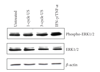
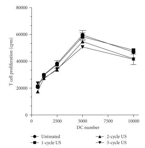
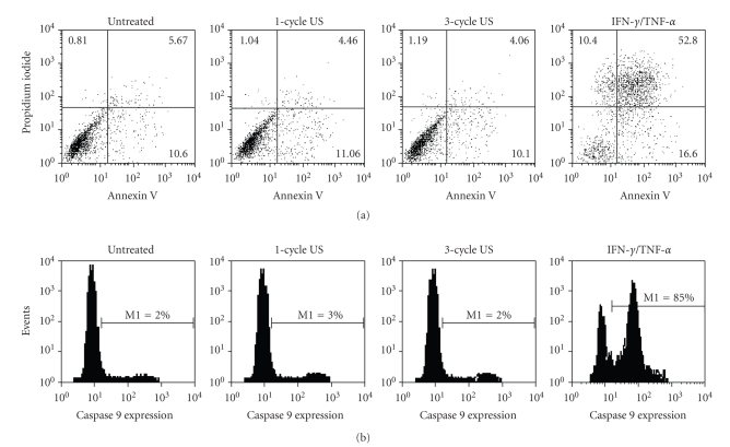
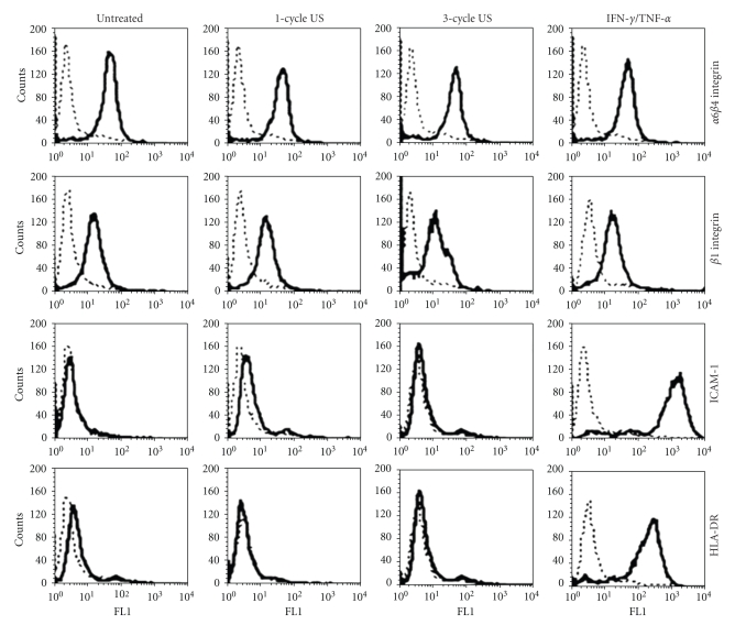
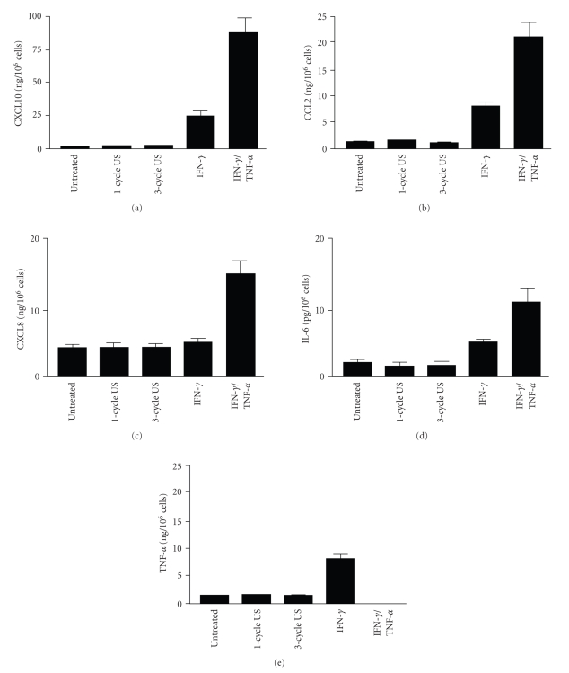
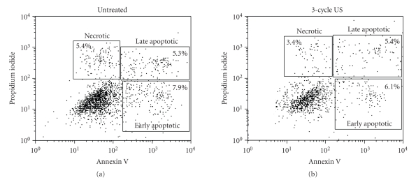
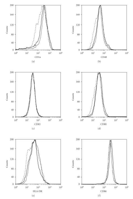
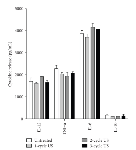
Low-frequency ultrasounds (US) are used to enhance drug transdermal transport. Although this phenomenon has been extensively analyzed, information on US effects on the single skin cell components is limited. Here, we investigated the possible effects of low-frequency US on viability and immune functions of cultured human keratinocytes and dendritic cells (DC), skin cells involved in the regulation of many immune-mediated dermatoses. We demonstrated that US, employed at low-frequency (42 KHz) and low-intensity (0.15 W/cm(2)) values known to enhance drug and water transdermal transport, did not affect extracellular-signal-regulated-kinase (ERK)1/2 activation, cell viability, or expression of adhesion molecules in cultured keratinocytes. Moreover, US at these work frequency and intensity did not influence the keratinocyte expression and release of immunomodulatory molecules. Similarly, cultured DC treated with low-frequency low-intensity US were viable, and did not show an altered membrane phenotype, cytokine profile, nor antigen presentation ability. However, intensity enhancement of low-frequency US to 5 W/cm(2) determined an increase of the apoptotic rate of both keratinocytes and DC as well as keratinocyte CXCL8 release and ERK1/2 activation, and DC CD40 expression. Our study sustains the employment of low-frequency and low-intensity US for treatment of those immune skin disorders, where keratinocytes and DC have a pathogenetic role.
低频超声(US)被用于增强药物经皮转运。尽管这一现象已得到广泛分析,但关于超声对单个皮肤细胞成分影响的信息有限。在此,我们研究了低频超声对培养的人角质形成细胞和树突状细胞(DC)活力及免疫功能的可能影响,角质形成细胞和树突状细胞是参与多种免疫介导性皮肤病调节的皮肤细胞。我们证明,以已知可增强药物和水经皮转运的低频(42千赫)和低强度(0.15瓦/平方厘米)使用超声,不会影响培养的角质形成细胞中细胞外信号调节激酶(ERK)1/2的激活、细胞活力或黏附分子的表达。此外,在这些工作频率和强度下的超声不会影响角质形成细胞免疫调节分子的表达和释放。同样,用低频低强度超声处理的培养DC具有活力,且未显示出膜表型、细胞因子谱或抗原呈递能力的改变。然而,将低频超声强度提高到5瓦/平方厘米会导致角质形成细胞和DC的凋亡率增加,以及角质形成细胞CXCL8释放和ERK1/2激活,和DC CD40表达增加。我们的研究支持在角质形成细胞和DC具有致病作用的免疫性皮肤病治疗中使用低频和低强度超声。