Lee Christopher Seungkyu, Kim Do Kyung, Lee Sung Chul
Department of Ophthalmology, Institute of Vision Research, Yonsei University College of Medicine, Seoul, Korea.
Korean J Ophthalmol. 2010 Feb;24(1):44-6. doi: 10.3341/kjo.2010.24.1.44. Epub 2010 Feb 5.
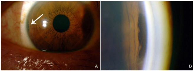
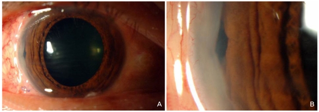
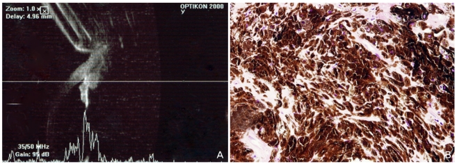
We report a case of ciliary body melanocytoma in a Korean patient, which presented as an intermittently painful pigmented iris mass and was successfully managed by iridocyclectomy. A 52-year-old healthy man presented with an irregularly-shaped and heavily-pigmented mass at the iris root of his right eye. Visual acuity of the right eye was 20/20 with normal intraocular pressure. Ultrasound biomicroscopy showed a 1.5x1.3-mm ciliary-body mass with extension into the iris root. Iridocyclectomy with scleral resection under a lamellar scleral flap was performed, and the histopathologic features of the resected tissue were consistent with melanocytoma of the ciliary body. The patient's visual acuity remained 20/20 with good postoperative cosmesis. During one year of follow-up, no signs of tumor recurrence were seen, and the patient reported resolution of the intermittent ocular pain in the involved eye.
我们报告了一例韩国患者的睫状体黑素细胞瘤,该肿瘤表现为间歇性疼痛的色素性虹膜肿物,并通过虹膜睫状体切除术成功治疗。一名52岁的健康男性,右眼虹膜根部出现一个形状不规则且色素沉着严重的肿物。右眼视力为20/20,眼压正常。超声生物显微镜检查显示一个1.5×1.3毫米的睫状体肿物,延伸至虹膜根部。在板层巩膜瓣下进行了巩膜切除术的虹膜睫状体切除术,切除组织的组织病理学特征与睫状体黑素细胞瘤一致。患者术后视力仍为20/20,美容效果良好,并在术后随访一年中未发现肿瘤复发迹象,患者自述患眼间歇性眼痛症状消失。