University of Lyon, Faculty of Medicine, EA 4170 Laboratory of Free Radicals, Energy Substrates and Cerebral Physiopathology, & Neurochem platform, Lyon, France.
PLoS One. 2010 Feb 16;5(2):e9211. doi: 10.1371/journal.pone.0009211.
The implication of nitric oxide (NO) in the development of human African trypanosomiasis (HAT) using an animal model, was examined. The manner by which the trypanocidal activity of NO is impaired in the periphery and in the brain of rats infected with Trypanosoma brucei brucei (T. b. brucei) was analyzed through: (i) the changes occurring in NO concentration in both peripheral (blood) and cerebral compartments; (ii) the activity of nNOS and iNOS enzymes; (iii) identification of the brain cell types in which the NO-pathways are particularly active during the time-course of the infection.
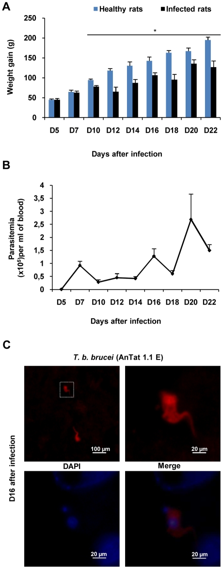
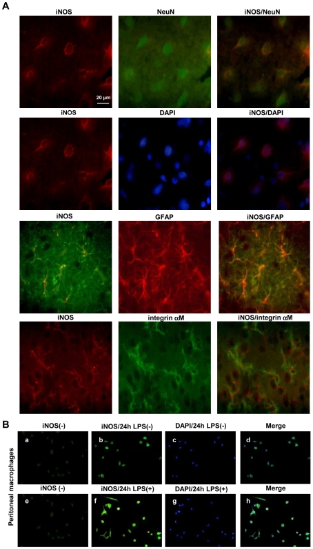
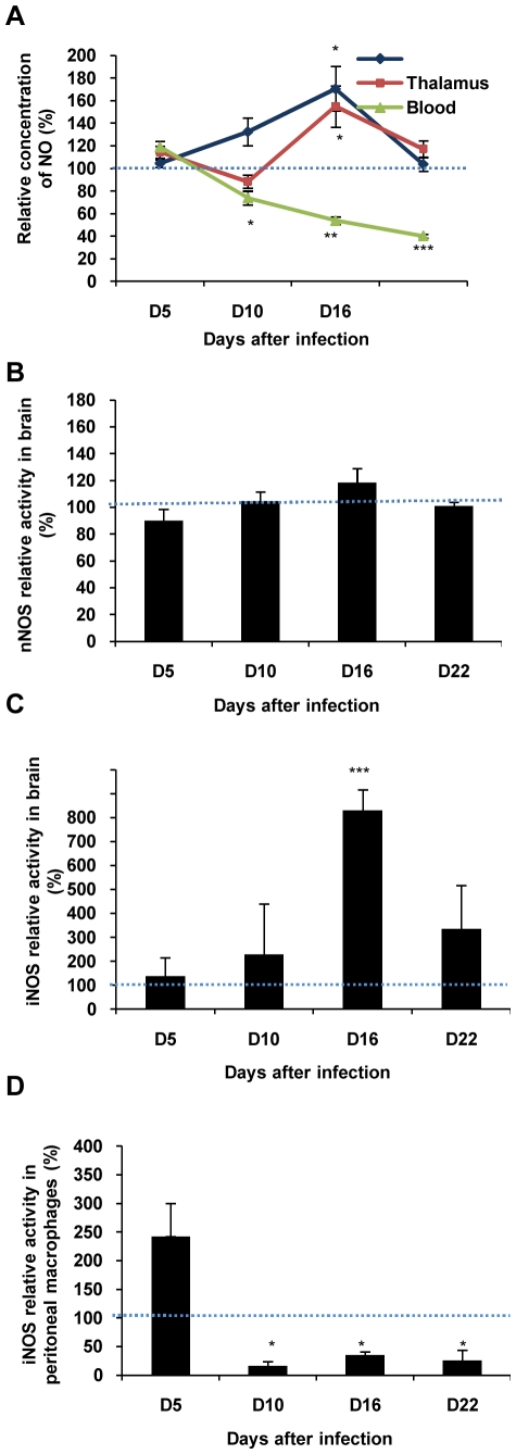
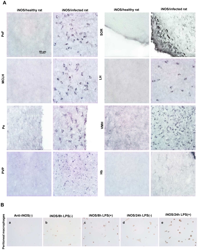
METHODOLOGY/PRINCIPAL FINDINGS: NO concentration (direct measures by voltammetry) was determined in central (brain) and peripheral (blood) compartments in healthy and infected animals at various days post-infection: D5, D10, D16 and D22. Opposite changes were observed in the two compartments. NO production increased in the brain (hypothalamus) from D10 (+32%) to D16 (+71%), but decreased in the blood from D10 (-22%) to D16 (-46%) and D22 (-60%). In parallel with NO measures, cerebral iNOS activity increased and peaked significantly at D16 (up to +700%). However, nNOS activity did not vary. Immunohistochemical staining confirmed iNOS activation in several brain regions, particularly in the hypothalamus. In peritoneal macrophages, iNOS activity decreased from D10 (-83%) to D16 (-65%) and D22 (-74%) similarly to circulating NO.
CONCLUSION/SIGNIFICANCE: The NO changes observed in our rat model were dependent on iNOS activity in both peripheral and central compartments. In the periphery, the NO production decrease may reflect an arginase-mediated synthesis of polyamines necessary to trypanosome growth. In the brain, the increased NO concentration may result from an enhanced activity of iNOS present in neurons and glial cells. It may be regarded as a marker of deleterious inflammatory reactions.
利用动物模型研究了一氧化氮(NO)在人类非洲锥虫病(HAT)发展中的作用。通过以下方式分析了 NO 杀锥虫活性在外周和感染布氏锥虫布鲁斯(T. b. brucei)的大鼠大脑中的受损方式:(i)外周(血液)和中枢(脑)隔室中 NO 浓度的变化;(ii)nNOS 和 iNOS 酶的活性;(iii)在感染过程的时间进程中,鉴定特别活跃的 NO 途径的脑细胞类型。
方法/主要发现:在感染后不同天数(D5、D10、D16 和 D22),通过伏安法直接测定健康和感染动物中枢(大脑)和外周(血液)隔室中的 NO 浓度。两个隔室观察到相反的变化。NO 产量从 D10 开始在大脑(下丘脑)中增加(+32%)到 D16(+71%),但从 D10 到 D16(-22%)和 D22(-46%)和 D22(-60%)在血液中减少。与 NO 测量同时,脑 iNOS 活性增加,在 D16 时显著增加(高达+700%)。然而,nNOS 活性没有变化。免疫组织化学染色证实 iNOS 在几个大脑区域(特别是在下丘脑)中被激活。在腹腔巨噬细胞中,iNOS 活性从 D10 开始减少(-83%)到 D16(-65%)和 D22(-74%),与循环 NO 相似。
结论/意义:我们的大鼠模型中观察到的 NO 变化取决于外周和中枢隔室中 iNOS 的活性。在外周,NO 产生的减少可能反映了必需的多胺的精氨酸酶介导的合成,以促进锥虫的生长。在大脑中,NO 浓度的增加可能是由于神经元和神经胶质细胞中 iNOS 活性增强所致。它可以被视为有害炎症反应的标志物。