• 文献检索
  • 文档翻译
  • 深度研究
  • 学术资讯
  • Suppr Zotero 插件Zotero 插件
  • 邀请有礼
  • 套餐&价格
  • 历史记录
应用&插件
Suppr Zotero 插件Zotero 插件浏览器插件Mac 客户端Windows 客户端微信小程序
定价
高级版会员购买积分包购买API积分包
服务
文献检索文档翻译深度研究API 文档MCP 服务
关于我们
关于 Suppr公司介绍联系我们用户协议隐私条款
关注我们

Suppr 超能文献

核心技术专利:CN118964589B侵权必究
粤ICP备2023148730 号-1Suppr @ 2026

文献检索

告别复杂PubMed语法,用中文像聊天一样搜索,搜遍4000万医学文献。AI智能推荐,让科研检索更轻松。

立即免费搜索

文件翻译

保留排版,准确专业,支持PDF/Word/PPT等文件格式,支持 12+语言互译。

免费翻译文档

深度研究

AI帮你快速写综述,25分钟生成高质量综述,智能提取关键信息,辅助科研写作。

立即免费体验

用于干细胞分化无创监测的光学光谱学。

Optical spectroscopy for noninvasive monitoring of stem cell differentiation.

作者信息

Downes Andrew, Mouras Rabah, Elfick Alistair

机构信息

School of Engineering, The University of Edinburgh, Edinburgh EH9 3JL, UK.

出版信息

J Biomed Biotechnol. 2010;2010:101864. doi: 10.1155/2010/101864. Epub 2010 Feb 16.

DOI:10.1155/2010/101864
PMID:20182537
原文链接:https://pmc.ncbi.nlm.nih.gov/articles/PMC2825551/
Abstract

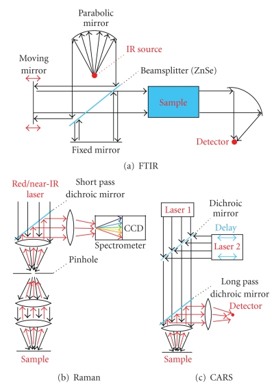
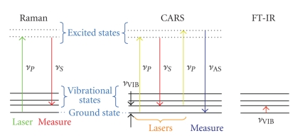
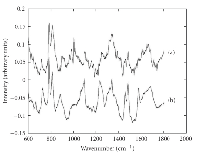
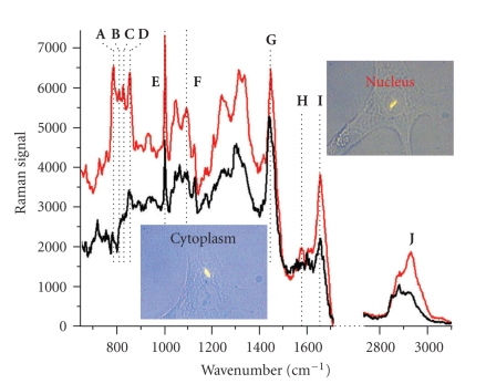
There is a requirement for a noninvasive technique to monitor stem cell differentiation. Several candidates based on optical spectroscopy are discussed in this review: Fourier transform infrared (FTIR) spectroscopy, Raman spectroscopy, and coherent anti-Stokes Raman scattering (CARS) microscopy. These techniques are briefly described, and the ability of each to distinguish undifferentiated from differentiated cells is discussed. FTIR spectroscopy has demonstrated its ability to distinguish between stem cells and their derivatives. Raman spectroscopy shows a clear reduction in DNA and RNA concentrations during embryonic stem cell differentiation (agreeing with the well-known reduction in the nucleus to cytoplasm ratio) and also shows clear increases in mineral content during differentiation of mesenchymal stem cells. CARS microscopy can map these DNA, RNA, and mineral concentrations at high speed, and Mutliplex CARS spectroscopy/microscopy is highlighted as the technique with most promise for future applications.

摘要

需要一种非侵入性技术来监测干细胞分化。本综述讨论了几种基于光谱学的候选技术:傅里叶变换红外(FTIR)光谱、拉曼光谱和相干反斯托克斯拉曼散射(CARS)显微镜。简要描述了这些技术,并讨论了每种技术区分未分化细胞和分化细胞的能力。FTIR光谱已证明其能够区分干细胞及其衍生物。拉曼光谱显示在胚胎干细胞分化过程中DNA和RNA浓度明显降低(与众所周知的细胞核与细胞质比例降低一致),并且在间充质干细胞分化过程中矿物质含量也明显增加。CARS显微镜可以高速绘制这些DNA、RNA和矿物质浓度图,而多重CARS光谱/显微镜被强调为最有前途的未来应用技术。

https://cdn.ncbi.nlm.nih.gov/pmc/blobs/c9b0/2825551/76fb7fc3f504/JBB2010-101864.006.jpg
https://cdn.ncbi.nlm.nih.gov/pmc/blobs/c9b0/2825551/8ea4495b0fd9/JBB2010-101864.001.jpg
https://cdn.ncbi.nlm.nih.gov/pmc/blobs/c9b0/2825551/c64f6aaabb3f/JBB2010-101864.002.jpg
https://cdn.ncbi.nlm.nih.gov/pmc/blobs/c9b0/2825551/f81ceb3a79c7/JBB2010-101864.003.jpg
https://cdn.ncbi.nlm.nih.gov/pmc/blobs/c9b0/2825551/b6e112277ba7/JBB2010-101864.004.jpg
https://cdn.ncbi.nlm.nih.gov/pmc/blobs/c9b0/2825551/4c5393af0801/JBB2010-101864.005.jpg
https://cdn.ncbi.nlm.nih.gov/pmc/blobs/c9b0/2825551/76fb7fc3f504/JBB2010-101864.006.jpg

相似文献

1
Optical spectroscopy for noninvasive monitoring of stem cell differentiation.用于干细胞分化无创监测的光学光谱学。
J Biomed Biotechnol. 2010;2010:101864. doi: 10.1155/2010/101864. Epub 2010 Feb 16.
2
Vibrational spectroscopy differentiates between multipotent and pluripotent stem cells.振动光谱可区分多能干细胞和多能干细胞。
Analyst. 2010 Dec;135(12):3126-32. doi: 10.1039/c0an00525h. Epub 2010 Oct 18.
3
Vibrational spectroscopy in stem cell characterisation: is there a niche?干细胞特性的振动光谱学:是否有一席之地?
Trends Biotechnol. 2014 May;32(5):254-62. doi: 10.1016/j.tibtech.2014.03.002. Epub 2014 Apr 3.
4
Chemical imaging with Fourier transform coherent anti-Stokes Raman scattering microscopy.傅里叶变换相干反斯托克斯拉曼散射显微镜的化学成像
Appl Opt. 2008 Nov 1;47(31):5790-8. doi: 10.1364/ao.47.005790.
5
Raman spectroscopy and coherent anti-Stokes Raman scattering imaging: prospective tools for monitoring skeletal cells and skeletal regeneration.拉曼光谱与相干反斯托克斯拉曼散射成像:监测骨骼细胞与骨骼再生的前瞻性工具。
J R Soc Interface. 2016 May;13(118). doi: 10.1098/rsif.2016.0182.
6
Adipose-derived mesenchymal stem cells' adipogenesis chemistry analyzed by FTIR and Raman metrics.采用傅里叶变换红外光谱和拉曼指标分析脂肪来源间充质干细胞的成脂化学。
J Lipid Res. 2024 Jul;65(7):100573. doi: 10.1016/j.jlr.2024.100573. Epub 2024 Jun 4.
7
Coherent transfer function of Fourier transform spectral interferometric coherent anti-Stokes Raman scattering microscopy.傅里叶变换光谱干涉相干反斯托克斯拉曼散射显微镜的相干传递函数。
J Opt Soc Am A Opt Image Sci Vis. 2011 Aug 1;28(8):1689-94. doi: 10.1364/JOSAA.28.001689.
8
Vibrational Spectroscopy for In Vitro Monitoring Stem Cell Differentiation.基于振动光谱的体外监测干细胞分化
Molecules. 2020 Nov 26;25(23):5554. doi: 10.3390/molecules25235554.
9
Label-free nonlinear optical microscopy detects early markers for osteogenic differentiation of human stem cells.无标记非线性光学显微镜检测人干细胞成骨分化的早期标志物。
Sci Rep. 2016 May 26;6:26716. doi: 10.1038/srep26716.
10
Probing primary mesenchymal stem cells differentiation status by micro-Raman spectroscopy.通过微拉曼光谱法探测原代间充质干细胞分化状态。
Spectrochim Acta A Mol Biomol Spectrosc. 2019 Apr 15;213:384-390. doi: 10.1016/j.saa.2019.01.069. Epub 2019 Jan 29.

引用本文的文献

1
Unlocking the Potential of Silver Nanoparticles: From Synthesis to Versatile Bio-Applications.释放银纳米颗粒的潜力:从合成到多样的生物应用
Pharmaceutics. 2024 Sep 21;16(9):1232. doi: 10.3390/pharmaceutics16091232.
2
Recent Advances in Monitoring Stem Cell Status and Differentiation Using Nano-Biosensing Technologies.利用纳米生物传感技术监测干细胞状态与分化的最新进展
Nanomaterials (Basel). 2022 Aug 25;12(17):2934. doi: 10.3390/nano12172934.
3
Multimodal Label-Free Monitoring of Adipogenic Stem Cell Differentiation Using Endogenous Optical Biomarkers.

本文引用的文献

1
The history and current status of fourier transform spectroscopy.傅里叶变换光谱学的历史与现状。
Appl Opt. 1966 May 1;5(5):845-54. doi: 10.1364/AO.5.000845.
2
Comparative materials differences revealed in engineered bone as a function of cell-specific differentiation.作为细胞特异性分化的函数,工程骨中揭示的比较材料差异。
Nat Mater. 2009 Sep;8(9):763-70. doi: 10.1038/nmat2505. Epub 2009 Jul 26.
3
A rapid, cost-effective method for counting human embryonic stem cell numbers as clumps.一种快速、经济高效的将人类胚胎干细胞集落作为团块进行计数的方法。
利用内源性光学生物标志物对脂肪生成干细胞分化进行多模态无标记监测。
Adv Funct Mater. 2021 Oct 20;31(43). doi: 10.1002/adfm.202103955. Epub 2021 Aug 6.
4
Metabolomic Alterations Do Not Induce Metabolic Burden in the Industrial Yeast M2n[pBKD2-]-C1 Engineered by Multiple δ-Integration of a Fungal β-Glucosidase Gene.代谢组学改变不会在通过真菌β-葡萄糖苷酶基因的多次δ整合工程改造的工业酵母M2n[pBKD2-]-C1中引发代谢负担。
Front Bioeng Biotechnol. 2019 Nov 28;7:376. doi: 10.3389/fbioe.2019.00376. eCollection 2019.
5
Nanobiosensing Platforms for Real-Time and Non-Invasive Monitoring of Stem Cell Pluripotency and Differentiation.用于实时、非侵入式监测干细胞多能性和分化的纳米生物传感平台。
Sensors (Basel). 2018 Aug 21;18(9):2755. doi: 10.3390/s18092755.
6
Single-Cell Analysis Using Hyperspectral Imaging Modalities.使用高光谱成像模式的单细胞分析
J Biomech Eng. 2018 Feb 1;140(2):0208021-02080216. doi: 10.1115/1.4038638.
7
FTIR Spectroscopic and Molecular Analysis during Differentiation of Pluripotent Stem Cells to Pancreatic Cells.多能干细胞向胰腺细胞分化过程中的傅里叶变换红外光谱和分子分析
Stem Cells Int. 2016;2016:6709714. doi: 10.1155/2016/6709714. Epub 2016 Aug 29.
8
Detection of early stage changes associated with adipogenesis using Raman spectroscopy under aseptic conditions.在无菌条件下使用拉曼光谱检测与脂肪生成相关的早期变化。
Cytometry A. 2015 Nov;87(11):1012-9. doi: 10.1002/cyto.a.22777. Epub 2015 Oct 6.
9
Fourier transform infrared microspectroscopy monitoring of 5-fluorouracil-induced apoptosis in SW620 colon cancer cells.傅里叶变换红外显微光谱法监测5-氟尿嘧啶诱导SW620结肠癌细胞凋亡
Mol Med Rep. 2015 Apr;11(4):2585-91. doi: 10.3892/mmr.2014.3088. Epub 2014 Dec 12.
10
Histologic and spectroscopic study of pluripotent stem cells after implant in ocular traumatic injuries in a murine model.小鼠模型中眼外伤植入多能干细胞后的组织学和光谱学研究。
Stem Cell Res Ther. 2014 Oct 20;5(5):119. doi: 10.1186/scrt509.
Biotechniques. 2009 Jul;47(1):599-606. doi: 10.2144/000113151.
4
Molecular identification by generating coherence between molecular normal modes using stimulated Raman scattering.通过受激拉曼散射在分子简正模式之间产生相干性来进行分子识别。
Opt Lett. 2009 Jun 15;34(12):1756-8. doi: 10.1364/ol.34.001756.
5
In-vivo multi-nonlinear optical imaging of a living cell using a supercontinuum light source generated from a photonic crystal fiber.使用由光子晶体光纤产生的超连续光源对活细胞进行体内多非线性光学成像。
Opt Express. 2006 Apr 3;14(7):2798-804. doi: 10.1364/oe.14.002798.
6
High-speed vibrational imaging and spectral analysis of lipid bodies by compound Raman microscopy.通过复合拉曼显微镜对脂质体进行高速振动成像和光谱分析。
J Phys Chem B. 2009 May 28;113(21):7681-6. doi: 10.1021/jp902231y.
7
Tracking the cell hierarchy in the human intestine using biochemical signatures derived by mid-infrared microspectroscopy.利用中红外光谱衍生的生化特征追踪人类肠道中的细胞层级结构。
Stem Cell Res. 2009 Jul;3(1):15-27. doi: 10.1016/j.scr.2009.02.003. Epub 2009 Feb 21.
8
Chemical imaging of live cancer cells in the natural aqueous environment.天然水环境中活癌细胞的化学成像。
Appl Spectrosc. 2009 Feb;63(2):164-71. doi: 10.1366/000370209787391969.
9
Label-free separation of human embryonic stem cells and their cardiac derivatives using Raman spectroscopy.利用拉曼光谱对人胚胎干细胞及其心脏衍生物进行无标记分离。
Anal Chem. 2009 Feb 15;81(4):1324-31. doi: 10.1021/ac801665m.
10
Label-free biomedical imaging with high sensitivity by stimulated Raman scattering microscopy.基于受激拉曼散射显微镜的高灵敏度无标记生物医学成像。
Science. 2008 Dec 19;322(5909):1857-61. doi: 10.1126/science.1165758.