• 文献检索
  • 文档翻译
  • 深度研究
  • 学术资讯
  • Suppr Zotero 插件Zotero 插件
  • 邀请有礼
  • 套餐&价格
  • 历史记录
应用&插件
Suppr Zotero 插件Zotero 插件浏览器插件Mac 客户端Windows 客户端微信小程序
定价
高级版会员购买积分包购买API积分包
服务
文献检索文档翻译深度研究API 文档MCP 服务
关于我们
关于 Suppr公司介绍联系我们用户协议隐私条款
关注我们

Suppr 超能文献

核心技术专利:CN118964589B侵权必究
粤ICP备2023148730 号-1Suppr @ 2026

文献检索

告别复杂PubMed语法,用中文像聊天一样搜索,搜遍4000万医学文献。AI智能推荐,让科研检索更轻松。

立即免费搜索

文件翻译

保留排版,准确专业,支持PDF/Word/PPT等文件格式,支持 12+语言互译。

免费翻译文档

深度研究

AI帮你快速写综述,25分钟生成高质量综述,智能提取关键信息,辅助科研写作。

立即免费体验

结肠癌相关结肠并发症的 CT 表现

CT findings of colonic complications associated with colon cancer.

机构信息

Department of Radiology, Cheonan Hospital, Soonchunhyang University, Cheonan 330-720, Korea.

出版信息

Korean J Radiol. 2010 Mar-Apr;11(2):211-21. doi: 10.3348/kjr.2010.11.2.211. Epub 2010 Feb 22.

DOI:10.3348/kjr.2010.11.2.211
PMID:20191069
原文链接:https://pmc.ncbi.nlm.nih.gov/articles/PMC2827785/
Abstract

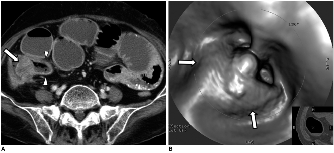
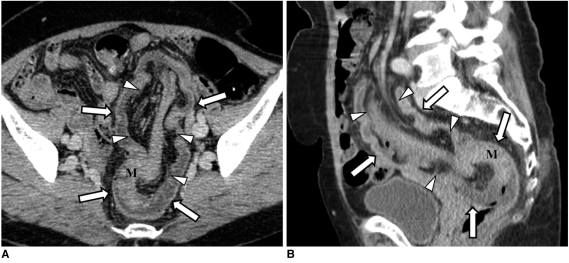
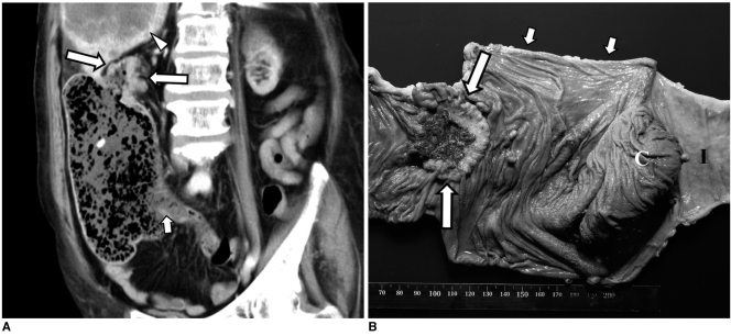
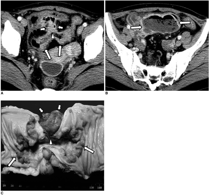
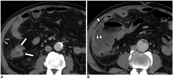
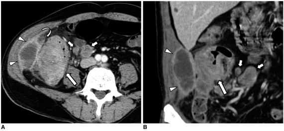
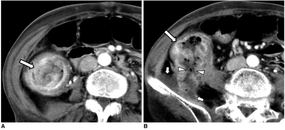
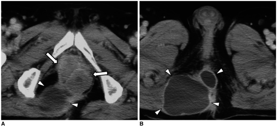
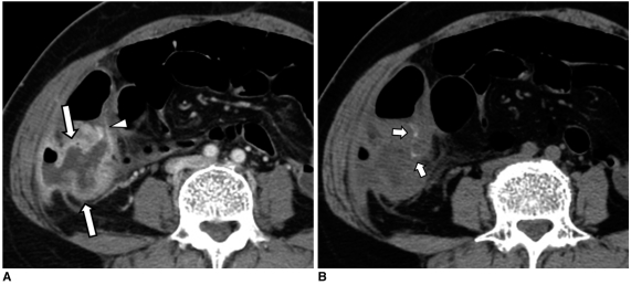
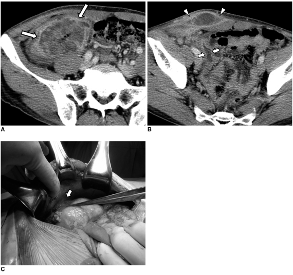
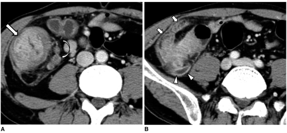
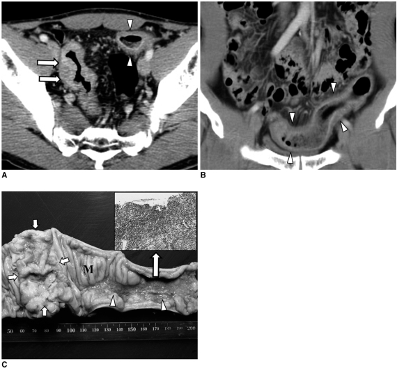
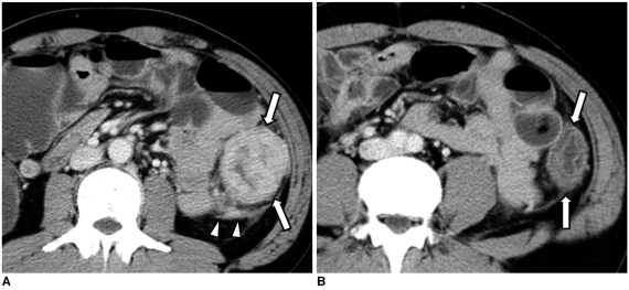
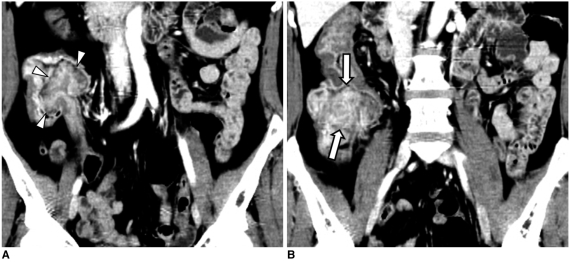
A broad spectrum of colonic complications can occur in patients with colon cancer. Clinically, some of these complications can obscure the presence of underlying malignancies in the colon and these complications may require emergency surgical management. The complications of the colon that can be associated with colon cancer include obstruction, perforation, abscess formation, acute appendicitis, ischemic colitis and intussusception. Although the majority of these complications only rarely occur, familiarity with the various manifestations of colon cancer complications will facilitate making an accurate diagnosis and administering prompt management in these situations. The purpose of this pictorial essay is to review the CT appearance of the colonic complications associated with colon cancer.

摘要

结直肠癌患者可能会出现广泛的结肠并发症。临床上,这些并发症中的一些可能会掩盖结肠中潜在的恶性肿瘤,这些并发症可能需要紧急手术治疗。可能与结直肠癌相关的结肠并发症包括梗阻、穿孔、脓肿形成、急性阑尾炎、缺血性结肠炎和肠套叠。虽然这些并发症大多数很少见,但熟悉结直肠癌并发症的各种表现形式将有助于在这些情况下做出准确的诊断并进行及时的治疗。本文旨在通过 CT 影像回顾结直肠癌相关结肠并发症的表现。

https://cdn.ncbi.nlm.nih.gov/pmc/blobs/729e/2827785/86b9e9b6d1db/kjr-11-211-g014.jpg
https://cdn.ncbi.nlm.nih.gov/pmc/blobs/729e/2827785/8674f7be0c80/kjr-11-211-g001.jpg
https://cdn.ncbi.nlm.nih.gov/pmc/blobs/729e/2827785/7df940ba55b0/kjr-11-211-g002.jpg
https://cdn.ncbi.nlm.nih.gov/pmc/blobs/729e/2827785/88bdab2a4171/kjr-11-211-g003.jpg
https://cdn.ncbi.nlm.nih.gov/pmc/blobs/729e/2827785/b69cfddf2fa1/kjr-11-211-g004.jpg
https://cdn.ncbi.nlm.nih.gov/pmc/blobs/729e/2827785/5e23609ffc07/kjr-11-211-g005.jpg
https://cdn.ncbi.nlm.nih.gov/pmc/blobs/729e/2827785/7371b9728a32/kjr-11-211-g006.jpg
https://cdn.ncbi.nlm.nih.gov/pmc/blobs/729e/2827785/4fa95d4d28d5/kjr-11-211-g007.jpg
https://cdn.ncbi.nlm.nih.gov/pmc/blobs/729e/2827785/2050d3fcb051/kjr-11-211-g008.jpg
https://cdn.ncbi.nlm.nih.gov/pmc/blobs/729e/2827785/1df9035a595f/kjr-11-211-g009.jpg
https://cdn.ncbi.nlm.nih.gov/pmc/blobs/729e/2827785/446c58165f9c/kjr-11-211-g010.jpg
https://cdn.ncbi.nlm.nih.gov/pmc/blobs/729e/2827785/d217ffdf1057/kjr-11-211-g011.jpg
https://cdn.ncbi.nlm.nih.gov/pmc/blobs/729e/2827785/8ad25098f0e7/kjr-11-211-g012.jpg
https://cdn.ncbi.nlm.nih.gov/pmc/blobs/729e/2827785/952d1cb0cda4/kjr-11-211-g013.jpg
https://cdn.ncbi.nlm.nih.gov/pmc/blobs/729e/2827785/86b9e9b6d1db/kjr-11-211-g014.jpg

相似文献

1
CT findings of colonic complications associated with colon cancer.结肠癌相关结肠并发症的 CT 表现
Korean J Radiol. 2010 Mar-Apr;11(2):211-21. doi: 10.3348/kjr.2010.11.2.211. Epub 2010 Feb 22.
2
[The contribution of ultrasonography to the diagnosis of acute intestinal diseases in adults].
JBR-BTR. 1998 Jun;81(3):144-9.
3
Transient colocolic intussusception.短暂性结肠结肠套叠
Clin Imaging. 2000 Jan-Feb;24(1):8-9. doi: 10.1016/s0899-7071(00)00154-6.
4
Ischemic origin of ulcerative colitis associated with potentially obstructing lesions of the colon.与结肠潜在梗阻性病变相关的溃疡性结肠炎的缺血性起源。
Radiology. 1972 Feb;102(2):249-52. doi: 10.1148/102.2.249.
5
CT evaluation of the acute abdomen: bowel pathology spectrum of disease.
Crit Rev Diagn Imaging. 1996 Aug;37(3):163-90.
6
Determining the site and causes of colonic obstruction with sonography.用超声检查确定结肠梗阻的部位和病因。
AJR Am J Roentgenol. 1994 Nov;163(5):1113-7. doi: 10.2214/ajr.163.5.7976885.
7
Giant colonic lipoma causing intussusception: CT scan and clinical findings.巨大结肠脂肪瘤致肠套叠:CT扫描及临床发现
Pan Afr Med J. 2019 Jan 16;32:27. doi: 10.11604/pamj.2019.32.27.18040. eCollection 2019.
8
Usefulness of CT in patients with ischemic colitis proximal to colonic cancer.CT在结肠癌近端缺血性结肠炎患者中的应用价值
AJR Am J Roentgenol. 1997 Apr;168(4):951-6. doi: 10.2214/ajr.168.4.9124147.
9
Spiral CT of colon cancer: imaging features and role in management.结肠癌的螺旋CT:影像学特征及其在治疗中的作用。
Radiographics. 2000 Mar-Apr;20(2):419-30. doi: 10.1148/radiographics.20.2.g00mc14419.
10
Ischemic colitis masquerading as colonic tumor: case report with review of literature.以结肠肿瘤为表现的缺血性结肠炎:病例报告并文献复习。
World J Gastroenterol. 2011 Dec 28;17(48):5324-6. doi: 10.3748/wjg.v17.i48.5324.

引用本文的文献

1
CT of acute abdomen in the elderly.老年人急腹症的CT检查
Insights Imaging. 2025 May 7;16(1):95. doi: 10.1186/s13244-025-01955-1.
2
DIVERT-Ca: unveiling the hidden link between acute diverticulitis and colorectal cancer risk-multicentre retrospective study.DIVERT-Ca研究:揭示急性憩室炎与结直肠癌风险之间的隐藏联系——多中心回顾性研究
Int J Colorectal Dis. 2025 Mar 15;40(1):68. doi: 10.1007/s00384-025-04858-1.
3
Integrated Pleiotropic Gene Set Unveils Comorbidity Insights across Digestive Cancers and Other Diseases.整合多效基因集揭示消化系统癌症及其他疾病的共病见解。

本文引用的文献

1
Gastrointestinal tract perforation: MDCT findings according to the perforation sites.胃肠道穿孔:根据穿孔部位的多层螺旋CT表现
Korean J Radiol. 2009 Jan-Feb;10(1):63-70. doi: 10.3348/kjr.2009.10.1.63.
2
Differences in patient postoperative and long-term outcomes between obstructive and perforated colonic cancer.梗阻性结肠癌与穿孔性结肠癌患者术后及长期预后的差异。
Am J Surg. 2008 Apr;195(4):427-32. doi: 10.1016/j.amjsurg.2007.02.027.
3
Colon cancer presenting as an appendiceal abscess in a young patient.一名年轻患者的结肠癌表现为阑尾脓肿。
Genes (Basel). 2024 Apr 10;15(4):478. doi: 10.3390/genes15040478.
4
Localization of Colorectal Cancer Lesions in Contrast-Computed Tomography Images via a Deep Learning Approach.通过深度学习方法对计算机断层扫描造影图像中的结直肠癌病变进行定位
Bioengineering (Basel). 2023 Aug 17;10(8):972. doi: 10.3390/bioengineering10080972.
5
The Neoplastic Side of the Abdominal Wall: A Comprehensive Pictorial Essay of Benign and Malignant Neoplasms.腹壁的肿瘤方面:良性和恶性肿瘤的综合图片论文
Diagnostics (Basel). 2023 Jan 15;13(2):315. doi: 10.3390/diagnostics13020315.
6
Interpretation discrepancies of abdominal imaging by on-call radiology residents: Evaluation of risk factors.值班放射科住院医师对腹部影像学的解读差异:危险因素评估。
PLoS One. 2022 Sep 9;17(9):e0274313. doi: 10.1371/journal.pone.0274313. eCollection 2022.
7
A Case of Acute Cerebral Infarction With Chief Complaints of Abdominal Pain and Bloody Diarrhoea: The Power of a Patient-Centered Inclusive Diagnostic Team.以腹痛和血性腹泻为主诉的急性脑梗死病例:以患者为中心的包容性诊断团队的力量
Cureus. 2022 Jul 28;14(7):e27386. doi: 10.7759/cureus.27386. eCollection 2022 Jul.
8
Automatic Detection and Segmentation of Colorectal Cancer with Deep Residual Convolutional Neural Network.基于深度残差卷积神经网络的结直肠癌自动检测与分割
Evid Based Complement Alternat Med. 2022 Mar 17;2022:3415603. doi: 10.1155/2022/3415603. eCollection 2022.
9
Aseptic abscess in the abdominal wall accompanied by monoclonal gammopathy simulating the local recurrence of rectal cancer: A case report.腹壁无菌性脓肿伴单克隆丙种球蛋白病,酷似直肠癌局部复发:一例报告
World J Clin Cases. 2022 Feb 16;10(5):1702-1708. doi: 10.12998/wjcc.v10.i5.1702.
10
Retroperitoneal Cecal Perforation Resulting from Obstructive Ascending Colon Adenocarcinoma.梗阻性升结肠癌导致的腹膜后盲肠穿孔
Case Rep Surg. 2020 Jan 6;2020:9371071. doi: 10.1155/2020/9371071. eCollection 2020.
Can J Surg. 2008 Feb;51(1):E15-6.
4
Adult intestinal intussusception: can abdominal MDCT distinguish an intussusception caused by a lead point?成人肠套叠:腹部多层螺旋CT能否鉴别由铅垂点引起的肠套叠?
Abdom Imaging. 2008 Sep-Oct;33(5):582-8. doi: 10.1007/s00261-007-9328-8.
5
A novel technique to relieve a closed-loop obstruction secondary to a competent ileocecal valve and an unresectable mid-colon tumor.一种用于缓解继发于回盲瓣功能正常和不可切除的结肠中段肿瘤的闭环梗阻的新技术。
J Gastrointest Surg. 2007 Oct;11(10):1365-7. doi: 10.1007/s11605-007-0230-z. Epub 2007 Jul 17.
6
Unusual abscesses associated with colon cancer: report of three cases.与结肠癌相关的罕见脓肿:三例报告
Acta Med Okayama. 2007 Apr;61(2):107-13. doi: 10.18926/AMO/32885.
7
Multi-detector row CT: spectrum of diseases involving the ileocecal area.多排探测器CT:累及回盲部的疾病谱
Radiographics. 2006 Sep-Oct;26(5):1373-90. doi: 10.1148/rg.265045191.
8
Adult intestinal intussusception: CT appearances and identification of a causative lead point.成人肠套叠:CT表现及病因引导点的识别
Radiographics. 2006 May-Jun;26(3):733-44. doi: 10.1148/rg.263055100.
9
Perforated colonic cancer presenting as intra-abdominal abscess.以腹腔内脓肿形式表现的穿孔性结肠癌
Int J Colorectal Dis. 2007 Jan;22(1):15-9. doi: 10.1007/s00384-006-0097-6. Epub 2006 Apr 20.
10
Abdominal wall and thigh abscess resulting from the penetration of ascending colon cancer.升结肠癌穿透导致腹壁及大腿脓肿
Acta Med Okayama. 2005 Dec;59(6):281-3. doi: 10.18926/AMO/31957.