Laganowski H C, Sherrard E S, Muir M G, Buckley R J
Moorfields Eye Hospital, London.
Br J Ophthalmol. 1991 Apr;75(4):212-6. doi: 10.1136/bjo.75.4.212.
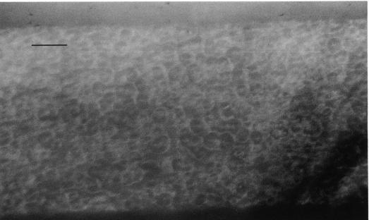
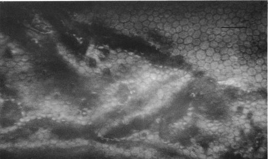
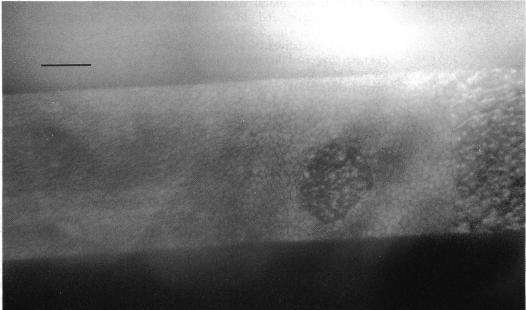
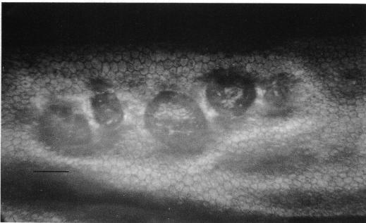
The literature suggests that posterior polymorphous dystrophy (PPD) may show features such as iridocorneal adhesions, glassy membranes, and pupillary ectropion which are typically ascribed to the iridocorneal endothelial (ICE) syndrome. This complicates diagnosis. PPD, unlike ICE, is familial, and ICE, unlike PPD, is usually progressive and frequently complicated by glaucoma: thus it is important to distinguish between them. To determine whether this could be achieved by specular microscopy, since the posterior corneal surface is abnormal in both conditions, 57 cases of ICE and 44 of PPD were repeatedly examined and photographed with the specular microscope. Progressive and/or static morphological features of the corneal endothelium and Descemet's membrane were found that were specific for each condition. Specular microscopy can thus provide a definitive diagnosis of ICE or PPD even in uncertain cases.
文献表明,后极性多形性营养不良(PPD)可能表现出虹膜角膜粘连、玻璃样膜和瞳孔外翻等特征,这些特征通常归因于虹膜角膜内皮(ICE)综合征。这使得诊断变得复杂。与ICE不同,PPD是家族性的,而与PPD不同,ICE通常是进行性的,并且经常并发青光眼:因此区分它们很重要。为了确定这是否可以通过镜面显微镜检查来实现,由于在这两种情况下角膜后表面均异常,因此对57例ICE患者和44例PPD患者进行了多次镜面显微镜检查并拍照。发现了角膜内皮和Descemet膜的进行性和/或静态形态学特征,这些特征对每种情况都是特异性的。因此,即使在不确定的病例中,镜面显微镜检查也可以对ICE或PPD做出明确诊断。