Department of Physiology and Pharmacology, Medical School, Southeast University, Nanjing 210009, People's Republic of China.
Int J Nanomedicine. 2010 Mar 9;5:109-16.
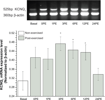
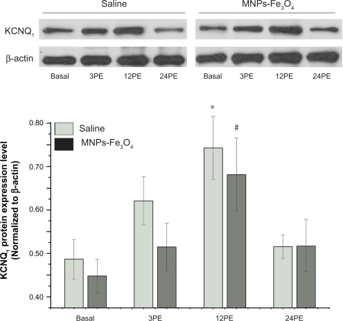
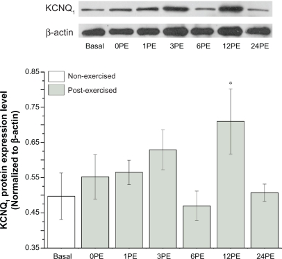
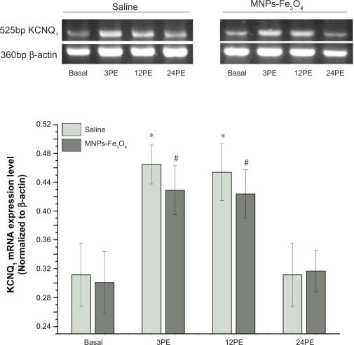
While the potential impact of magnetic nanoparticles (MNPs) has been widely explored in almost all medical fields, including cardiology, one question remains; that is whether MNPs interfere with cardiac physiological processes such as the expression and function of ion channels, especially in vivo. KCNQ(1) channels are richly expressed in cardiac myocytes and are critical to the repolarization of cardiac myocytes. In this study, we evaluated the effects of Fe(3)O(4)-magnetic nanoparticles (MNPs-Fe(3)O(4)) on the expression of KCNQ(1) in cardiac muscle of mice at rest and at different times following a single bout of swimming (SBS). Firstly, we demonstrated that the expression levels of KCNQ(1) channels are significantly up-regulated in mice following a SBS by means of reverse transcription polymerase chain reaction (RT-PCR) and western-blot. After treating mice with normal saline or pure MNPs-Fe(3)O(4) separately, we studied the potential effect of MNPs-Fe(3)O(4) on the expression profile of KCNQ(1) in mouse cardiac muscle following a SBS. A SBS increased the transcription of KCNQ(1) at 3 hours post exercise (3PE) 164% +/- 24% and at 12 hours post exercise (12PE) by 159% +/- 23% (P < 0.05), and up-regulated KCNQ(1) protein 161% +/- 27% at 12PE (P < 0.05) in saline mice. In MNPs-Fe(3)O(4) mice, KCNQ(1) mRNA increased by 151% +/- 14% and 147% +/- 12% at 3 and 12 PE, respectively (P <0.05). Meanwhile, an increase of 152% +/- 14% in KCNQ(1) protein was also detected at by 12PE. These results indicated that the administration of MNPs-Fe(3)O(4) did not cause any apparent effects on the expression profile of KCNQ(1) in rested or exercised mice cardiac muscle. Our studies suggest a novel path of KCNQ(1) current adaptations in the heart during physical exercise and in addition provide some useful information for the biomedical application of MNPs which are imperative to advance nanomedicine.
虽然磁性纳米粒子(MNPs)的潜在影响已在几乎所有医学领域得到广泛研究,包括心脏病学,但仍有一个问题悬而未决,即 MNPs 是否会干扰心脏的生理过程,如离子通道的表达和功能,特别是在体内。KCNQ(1)通道在心肌细胞中丰富表达,对心肌细胞的复极化至关重要。在这项研究中,我们评估了 Fe(3)O(4)-磁性纳米粒子(MNPs-Fe(3)O(4))对休息时和单次游泳后(SBS)不同时间点小鼠心肌中 KCNQ(1)表达的影响。首先,我们通过逆转录聚合酶链反应(RT-PCR)和蛋白质印迹证明 SBS 后小鼠的 KCNQ(1)通道表达水平显著上调。在分别用生理盐水或纯 MNPs-Fe(3)O(4)处理小鼠后,我们研究了 MNPs-Fe(3)O(4)对 SBS 后小鼠心肌中 KCNQ(1)表达谱的潜在影响。SBS 使运动后 3 小时(3PE)的 KCNQ(1)转录增加 164% +/- 24%,运动后 12 小时(12PE)增加 159% +/- 23%(P < 0.05),并使 KCNQ(1)蛋白在 12PE 时增加 161% +/- 27%(P < 0.05)在盐水小鼠中。在 MNPs-Fe(3)O(4)小鼠中,KCNQ(1)mRNA 在 3 和 12 PE 时分别增加 151% +/- 14%和 147% +/- 12%(P <0.05)。同时,在 12PE 时还检测到 KCNQ(1)蛋白增加 152% +/- 14%。这些结果表明,MNPs-Fe(3)O(4)的给药对休息或运动小鼠心肌中 KCNQ(1)的表达谱没有造成明显影响。我们的研究为运动过程中心脏 KCNQ(1)电流适应性提供了一条新途径,并为磁纳米粒子的生物医学应用提供了一些有用的信息,这对推进纳米医学至关重要。