Division of Rheumatology & Clinical Immunology, University of Florida, 1600 SW Archer Rd, Gainesville, FL 32610-0221, USA.
Arthritis Res Ther. 2010;12(3):R90. doi: 10.1186/ar3017. Epub 2010 May 18.
More than half of systemic lupus erythematosus (SLE) patients show evidence of excess type I interferon (IFN-I) production, a phenotype associated with renal disease and certain autoantibodies. However, detection of IFN-I proteins in serum is unreliable, and the measurement of interferon-stimulated gene (ISG) expression is expensive and time consuming. The aim of this study was to identify a surrogate marker for IFN-I activity in clinical samples for monitoring disease activity and response to therapy.
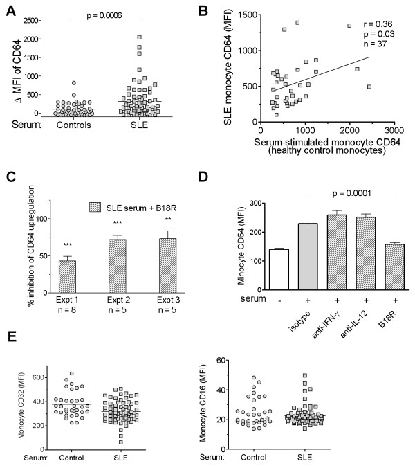
Monocyte surface expression of Fcgamma receptors (FcgammaRs), chemokine receptors, and activation markers were analyzed with flow cytometry in whole blood from patients with SLE and healthy controls. FcgammaR expression also was measured in peripheral blood mononuclear cells (PBMCs) from healthy controls cultured with Toll-like receptor (TLR) agonists, cytokines, or serum from SLE patients. Expression of ISGs was analyzed with real-time PCR.
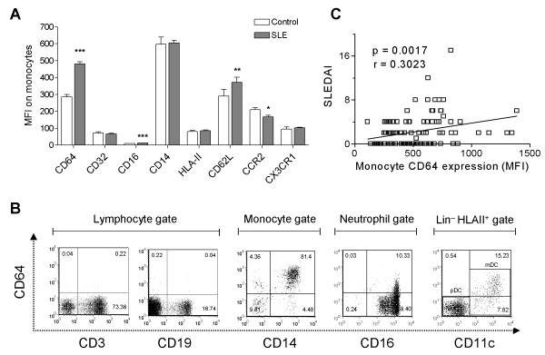
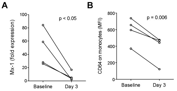
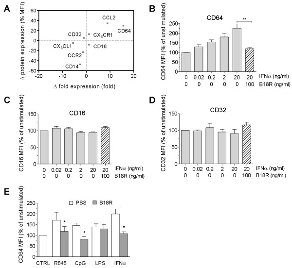
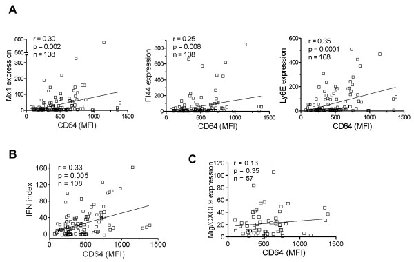
Circulating CD14+ monocytes from SLE patients showed increased surface expression of FcgammaRI (CD64). The mean fluorescent intensity of CD64 staining correlated highly with the ISG expression (MX1, IFI44, and Ly6E). In vitro, IFN-I as well as TLR7 and TLR9 agonists, induced CD64 expression on monocytes from healthy controls. Exposure of monocytes from healthy controls to SLE sera also upregulated the expression of CD64 in an IFN-I-dependent manner. Decreased CD64 expression was observed concomitant with the reduction of ISG expression after high-dose corticosteroid therapy.
Expression of CD64 on circulating monocytes is IFN-I inducible and highly correlated with ISG expression. Flow-cytometry analysis of CD64 expression on circulating monocytes is a convenient and rapid approach for estimating IFN-I levels in SLE patients.
超过一半的系统性红斑狼疮(SLE)患者表现出Ⅰ型干扰素(IFN-I)产生过多的证据,这种表型与肾脏疾病和某些自身抗体有关。然而,血清中 IFN-I 蛋白的检测不可靠,干扰素刺激基因(ISG)表达的测量既昂贵又耗时。本研究旨在寻找一种可替代的 IFN-I 活性的标志物,用于监测疾病的活动度和对治疗的反应。
用流式细胞术分析 SLE 患者和健康对照者全血中单核细胞表面 Fcγ 受体(FcγR)、趋化因子受体和激活标志物的表达。还测量了健康对照者外周血单个核细胞(PBMCs)在 TLR 激动剂、细胞因子或 SLE 患者血清培养物中 FcγR 的表达。用实时 PCR 分析 ISG 的表达。
SLE 患者循环 CD14+单核细胞表面 FcγRI(CD64)表达增加。CD64 染色的平均荧光强度与 ISG 表达(MX1、IFI44 和 Ly6E)高度相关。体外,IFN-I 以及 TLR7 和 TLR9 激动剂诱导健康对照者单核细胞 CD64 的表达。IFN-I 依赖性方式,健康对照者单核细胞暴露于 SLE 血清也上调 CD64 的表达。大剂量皮质类固醇治疗后,ISG 表达减少的同时,CD64 表达也减少。
循环单核细胞上 CD64 的表达可被 IFN-I 诱导,与 ISG 表达高度相关。流式细胞术分析循环单核细胞 CD64 的表达是一种估计 SLE 患者 IFN-I 水平的简便、快速方法。