Biopathology Department, Tor Vergata University, Rome, Italy.
J Transl Med. 2010 May 20;8:48. doi: 10.1186/1479-5876-8-48.
MicroRNAs are highly conserved, noncoding RNAs involved in post-transcriptional gene silencing. They have been shown to participate in a wide range of biological processes, including myogenesis and muscle regeneration. The goal of this study is to test the hypothesis that myo-miRs (myo = muscle + miR = miRNA) expression is altered in muscle from patients affected by myotonic dystrophy type 1 (DM1), the most frequently inherited neuromuscular disease in adults. In order to gain better insights about the role of miRNAs in the DM1 pathogenesis, we have also analyzed the muscular expression of miR-103 and miR-107, which have been identified in silico as attractive candidates for binding to the DMPK mRNA.
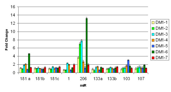
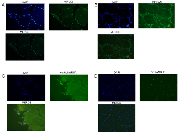
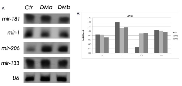
To this aim, we have profiled the expression of miR-133 (miR-133a, miR-133b), miR-1, miR-181 (miR-181a, miR-181b, miR-181c) and miR-206, that are specifically induced during myogenesis in cardiac and skeletal muscle tissues. miR-103 and miR-107, highly expressed in brain, heart and muscle have also been included in this study. QRT-PCR experiments have been performed on RNA from vastus lateralis biopsies of DM1 patients (n = 7) and control subjects (n = 4). Results of miRNAs expression have been confirmed by Northern blot, whereas in situ hybridization technique have been performed to localize misexpressed miRNAs on muscle sections from DM1 and control individuals.
Only miR-206 showed an over-expression in 5 of 7 DM1 patients (threshold = 2, fold change between 1.20 and 13.22, average = 5.37) compared to the control group. This result has been further confirmed by Northern blot analysis (3.37-fold overexpression, R2 = 0.89). In situ hybridization localized miR-206 to nuclear site both in normal and DM1 tissues. Cellular distribution in DM1 tissues includes also the nuclear regions of centralized nuclei, with a strong signal corresponding to nuclear clumps.
This work provides, for the first time, evidences about miRNAs misexpression in DM1 muscle tissues, adding a new element in the pathogenesis of this complex genetic disease.
微小 RNA 是高度保守的非编码 RNA,参与转录后基因沉默。它们已被证明参与广泛的生物过程,包括肌生成和肌肉再生。本研究的目的是检验这样一个假设,即肌源性微小 RNA(miR-133、miR-1、miR-181 和 miR-206)的表达在患有 1 型肌强直性营养不良(DM1)的患者的肌肉中发生改变,DM1 是成人中最常见的遗传性神经肌肉疾病。为了更好地了解 miRNA 在 DM1 发病机制中的作用,我们还分析了肌内 miR-103 和 miR-107 的表达,这些 miRNA 已在计算机中被鉴定为与 DMPK mRNA 结合的有吸引力的候选物。
为此,我们对心脏和骨骼肌组织中肌生成特异性诱导的 miR-133(miR-133a、miR-133b)、miR-1、miR-181(miR-181a、miR-181b、miR-181c)和 miR-206 的表达进行了分析。本研究还包括在大脑、心脏和肌肉中高度表达的 miR-103 和 miR-107。对 7 名 DM1 患者(n=7)和 4 名对照组患者的股外侧肌活检组织中的 RNA 进行 QRT-PCR 实验。通过Northern blot 验证了 miRNA 表达结果,通过原位杂交技术将异常表达的 miRNA 定位在 DM1 和对照组个体的肌肉切片上。
与对照组相比,只有 miR-206 在 7 名 DM1 患者中的 5 名中表现出过度表达(阈值=2,倍数变化在 1.20 到 13.22 之间,平均值=5.37)。Northern blot 分析进一步证实了这一结果(3.37 倍过表达,R2=0.89)。原位杂交将 miR-206 定位在正常和 DM1 组织的核区。在 DM1 组织中,细胞分布还包括集中核的核区,有强烈的信号对应核团。
本研究首次提供了 DM1 肌肉组织中 miRNA 表达异常的证据,为这种复杂的遗传疾病的发病机制增加了一个新的元素。