Department of Neurology, Chang Gung Memorial Hospital-Kaohsiung Medical Center and Chang Gung University College of Medicine, Niaosung, Taiwan.
BMC Neurol. 2010 Jul 6;10:59. doi: 10.1186/1471-2377-10-59.
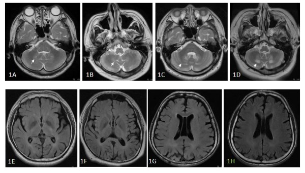
Cerebrotendinous xanthomatosis (CTX) is a rare genetic disorder. Recent studies show that brain damage in CTX patients extends beyond the abnormalities observed on conventional magnetic resonance imaging (MRI). We studied the MRI and 99 mTc-ethyl cysteinate dimer single photon emission computed tomography (SPECT) findings of CTX patients and made a correlation with the neuropsychological presentations.
Diffusion tensor imaging (DTI) and 3D T1-weighted images of five CTX patients were compared with 15 age-matched controls. Voxel-based morphometry (VBM) was use to delineate gray matter (GM) and white matter (WM) volume loss. Fractional anisotropy (FA), mean diffusivity (MD), and eigenvalues derived from DTI were used to detect WM changes and correlate with neuropsychological results. SPECT functional studies were used to correlate with GM changes.
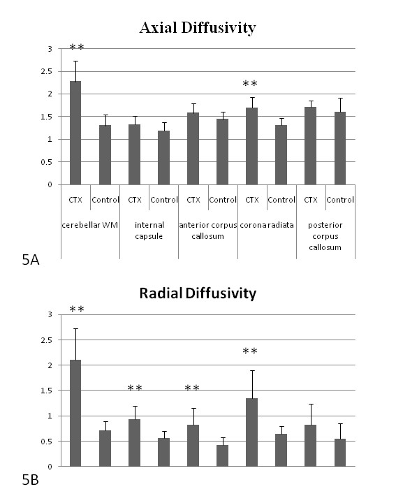
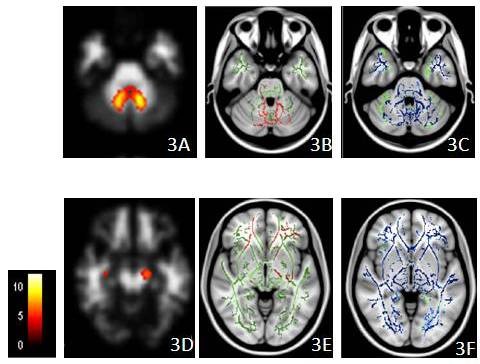
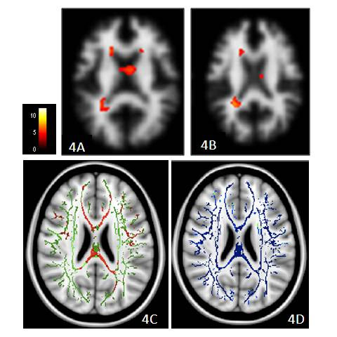
Cognitive results showed that aside from moderate mental retardation, the patient group performed worse in all cognitive domains. Despite the extensive GM atrophy pattern, the cerebellum, peri-Sylvian regions and parietal-occipital regions were correlated with SPECT results. WM atrophy located in the peri-dentate and left cerebral peduncle areas corresponded with changes in diffusion measures, while axial and radial diffusivity suggested both demyelinating and axonal changes. Changes in FA and MD were preceded by VBM in the corpus callosum and corona radiata. Cognitive results correlated with FA changes.
In CTX, GM atrophy affected the perfusion patterns. Changes in WM included atrophy, and axonal changes with demyelination. Disconnection of major fiber tracts among different cortical regions may contribute to cognitive impairment.
脑腱黄瘤病(CTX)是一种罕见的遗传性疾病。最近的研究表明,CTX 患者的脑损伤超出了常规磁共振成像(MRI)观察到的异常。我们研究了 CTX 患者的 MRI 和 99mTc-乙基半胱氨酸二聚体单光子发射计算机断层扫描(SPECT)结果,并与神经心理学表现进行了相关性分析。
比较了五例 CTX 患者的弥散张量成像(DTI)和三维 T1 加权图像与 15 名年龄匹配的对照组。使用基于体素的形态计量学(VBM)来描绘灰质(GM)和白质(WM)体积损失。各向异性分数(FA)、平均弥散度(MD)和 DTI 导出的特征值用于检测 WM 变化,并与神经心理学结果相关联。SPECT 功能研究用于与 GM 变化相关联。
认知结果表明,除了中度智力障碍外,患者组在所有认知领域的表现都较差。尽管 GM 萎缩模式广泛,但小脑、大脑外侧裂周围区域和顶枕叶区域与 SPECT 结果相关。位于齿状回周围和左侧大脑脚区域的 WM 萎缩与弥散测量的变化相对应,而轴向和径向弥散度提示脱髓鞘和轴突变化。胼胝体和放射冠的 VBM 先于 FA 和 MD 的变化。认知结果与 FA 变化相关。
在 CTX 中,GM 萎缩影响了灌注模式。WM 的变化包括萎缩和脱髓鞘的轴突变化。不同皮质区域之间主要纤维束的断开可能导致认知障碍。