• 文献检索
  • 文档翻译
  • 深度研究
  • 学术资讯
  • Suppr Zotero 插件Zotero 插件
  • 邀请有礼
  • 套餐&价格
  • 历史记录
应用&插件
Suppr Zotero 插件Zotero 插件浏览器插件Mac 客户端Windows 客户端微信小程序
定价
高级版会员购买积分包购买API积分包
服务
文献检索文档翻译深度研究API 文档MCP 服务
关于我们
关于 Suppr公司介绍联系我们用户协议隐私条款
关注我们

Suppr 超能文献

核心技术专利:CN118964589B侵权必究
粤ICP备2023148730 号-1Suppr @ 2026

文献检索

告别复杂PubMed语法,用中文像聊天一样搜索,搜遍4000万医学文献。AI智能推荐,让科研检索更轻松。

立即免费搜索

文件翻译

保留排版,准确专业,支持PDF/Word/PPT等文件格式,支持 12+语言互译。

免费翻译文档

深度研究

AI帮你快速写综述,25分钟生成高质量综述,智能提取关键信息,辅助科研写作。

立即免费体验

CT血管造影:当前技术与临床应用。

CT angiography: current technology and clinical use.

作者信息

Kumamaru Kanako K, Hoppel Bernice E, Mather Richard T, Rybicki Frank J

机构信息

Department of Radiology, University of Tokyo Hospital, Tokyo, Japan.

出版信息

Radiol Clin North Am. 2010 Mar;48(2):213-35, vii. doi: 10.1016/j.rcl.2010.02.006.

DOI:10.1016/j.rcl.2010.02.006
PMID:20609871
原文链接:https://pmc.ncbi.nlm.nih.gov/articles/PMC2901244/
Abstract

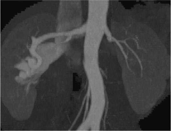
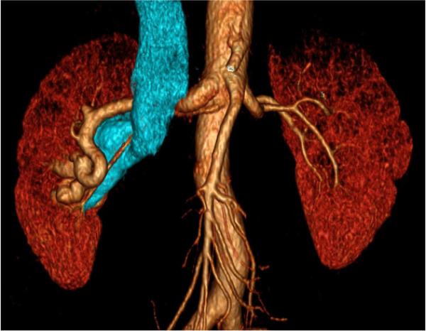
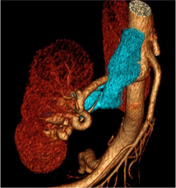
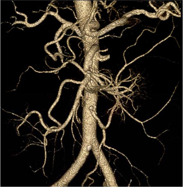
Since 1958, catheter angiography has assumed the role of gold standard for vascular imaging, despite the invasive nature of the procedure. Less invasive techniques for vascular imaging, such as computed tomographic angiography (CTA), have been developed and have matured in conjunction with developments in catheter arteriography. In a few cases, such as imaging, the aorta and the pulmonary arteries, CTA has supplanted catheter angiography as the gold standard. The expanding role of CTA emphasizes the need for deep, broad-based understanding of physical principles. This review describes CT hardware and associated software for angiography. The fundamentals of CTA physics are complemented with several clinical examples.

摘要

自1958年以来,尽管导管血管造影术具有侵入性,但它一直是血管成像的金标准。血管成像的侵入性较小的技术,如计算机断层血管造影(CTA),已随着导管动脉造影术的发展而得到开发并成熟。在少数情况下,如对主动脉和肺动脉进行成像时,CTA已取代导管血管造影术成为金标准。CTA作用的不断扩大凸显了深入、全面理解物理原理的必要性。本综述描述了用于血管造影的CT硬件及相关软件。CTA物理基础辅以若干临床实例。

https://cdn.ncbi.nlm.nih.gov/pmc/blobs/b012/2901244/b5995ca25480/nihms-180757-f0026.jpg
https://cdn.ncbi.nlm.nih.gov/pmc/blobs/b012/2901244/3efb262d8c4d/nihms-180757-f0001.jpg
https://cdn.ncbi.nlm.nih.gov/pmc/blobs/b012/2901244/d98689fe2819/nihms-180757-f0002.jpg
https://cdn.ncbi.nlm.nih.gov/pmc/blobs/b012/2901244/276a706046b3/nihms-180757-f0003.jpg
https://cdn.ncbi.nlm.nih.gov/pmc/blobs/b012/2901244/d8fec12e2a31/nihms-180757-f0004.jpg
https://cdn.ncbi.nlm.nih.gov/pmc/blobs/b012/2901244/415115c392bb/nihms-180757-f0005.jpg
https://cdn.ncbi.nlm.nih.gov/pmc/blobs/b012/2901244/826b10d10fb7/nihms-180757-f0006.jpg
https://cdn.ncbi.nlm.nih.gov/pmc/blobs/b012/2901244/f6a1dc6f6b5f/nihms-180757-f0007.jpg
https://cdn.ncbi.nlm.nih.gov/pmc/blobs/b012/2901244/e7189bad27a9/nihms-180757-f0008.jpg
https://cdn.ncbi.nlm.nih.gov/pmc/blobs/b012/2901244/4c65fa992fd3/nihms-180757-f0009.jpg
https://cdn.ncbi.nlm.nih.gov/pmc/blobs/b012/2901244/268b53ab77f3/nihms-180757-f0010.jpg
https://cdn.ncbi.nlm.nih.gov/pmc/blobs/b012/2901244/41646b74c567/nihms-180757-f0011.jpg
https://cdn.ncbi.nlm.nih.gov/pmc/blobs/b012/2901244/fc91e152959f/nihms-180757-f0012.jpg
https://cdn.ncbi.nlm.nih.gov/pmc/blobs/b012/2901244/77ee674c8313/nihms-180757-f0013.jpg
https://cdn.ncbi.nlm.nih.gov/pmc/blobs/b012/2901244/94a99d87e274/nihms-180757-f0014.jpg
https://cdn.ncbi.nlm.nih.gov/pmc/blobs/b012/2901244/2e9265ccfc6a/nihms-180757-f0015.jpg
https://cdn.ncbi.nlm.nih.gov/pmc/blobs/b012/2901244/327185bd4178/nihms-180757-f0016.jpg
https://cdn.ncbi.nlm.nih.gov/pmc/blobs/b012/2901244/653b00fd1d05/nihms-180757-f0017.jpg
https://cdn.ncbi.nlm.nih.gov/pmc/blobs/b012/2901244/e0097cae4a59/nihms-180757-f0018.jpg
https://cdn.ncbi.nlm.nih.gov/pmc/blobs/b012/2901244/09e4a315adae/nihms-180757-f0019.jpg
https://cdn.ncbi.nlm.nih.gov/pmc/blobs/b012/2901244/f9fcd007606f/nihms-180757-f0020.jpg
https://cdn.ncbi.nlm.nih.gov/pmc/blobs/b012/2901244/249e52e5d3e7/nihms-180757-f0021.jpg
https://cdn.ncbi.nlm.nih.gov/pmc/blobs/b012/2901244/4a00306e5922/nihms-180757-f0022.jpg
https://cdn.ncbi.nlm.nih.gov/pmc/blobs/b012/2901244/e69826f44f94/nihms-180757-f0023.jpg
https://cdn.ncbi.nlm.nih.gov/pmc/blobs/b012/2901244/52c42edd925b/nihms-180757-f0024.jpg
https://cdn.ncbi.nlm.nih.gov/pmc/blobs/b012/2901244/dfa92e60e9bb/nihms-180757-f0025.jpg
https://cdn.ncbi.nlm.nih.gov/pmc/blobs/b012/2901244/b5995ca25480/nihms-180757-f0026.jpg

相似文献

1
CT angiography: current technology and clinical use.CT血管造影:当前技术与临床应用。
Radiol Clin North Am. 2010 Mar;48(2):213-35, vii. doi: 10.1016/j.rcl.2010.02.006.
2
[The importance of multidetector computed tomography in the vascular imaging].[多层螺旋计算机断层扫描在血管成像中的重要性]
Orv Hetil. 2009 Jul 19;150(29):1351-60. doi: 10.1556/OH.2009.28664.
3
Three-dimensional spiral computed tomographic angiography: an alternative imaging modality for the abdominal aorta and its branches.三维螺旋计算机断层血管造影术:腹主动脉及其分支的一种替代成像方式。
J Vasc Surg. 1993 Oct;18(4):656-64; discussion 665.
4
3D fusion of coronary CT angiography and CT myocardial perfusion imaging: Intuitive assessment of morphology and function.冠状动脉 CT 血管造影与 CT 心肌灌注成像的三维融合:形态学与功能的直观评估。
J Cardiovasc Comput Tomogr. 2017 Nov;11(6):437-443. doi: 10.1016/j.jcct.2017.09.003. Epub 2017 Sep 5.
5
Spiral CT: vascular applications.螺旋CT:血管应用
Eur J Radiol. 1998 Aug;28(1):18-29. doi: 10.1016/s0720-048x(98)00009-6.
6
Multislice spiral computed tomography angiography of mesenteric arteries.肠系膜动脉的多层螺旋计算机断层血管造影术。
Lancet. 2001 Aug 25;358(9282):638-9. doi: 10.1016/S0140-6736(01)05786-5.
7
Review of CT angiography of aorta.主动脉CT血管造影术综述。
Radiol Clin North Am. 2007 May;45(3):461-83, viii. doi: 10.1016/j.rcl.2007.04.010.
8
Congenital anomalies of the aorta and vena cava: 16-detector-row CT imaging findings.主动脉和腔静脉先天性异常:16排CT成像表现
Diagn Interv Radiol. 2008 Sep;14(3):163-71.
9
Nonrigid registration-based coronary artery motion correction for cardiac computed tomography.基于非刚性配准的冠状动脉运动校正在心脏 CT 中的应用。
Med Phys. 2012 Jul;39(7):4245-54. doi: 10.1118/1.4725712.
10
Coronary 64-slice computed tomographic angiography models employing aortic root and selective catheter directed contrast enhancement in swine: technical feasibility and preliminary results using 3D and 4D reconstructions.猪冠状动脉64层计算机断层血管造影模型:采用主动脉根部和选择性导管引导对比剂增强技术,3D和4D重建的技术可行性及初步结果
Int J Cardiovasc Imaging. 2006 Jun-Aug;22(3-4):517-31. doi: 10.1007/s10554-006-9079-6. Epub 2006 Mar 15.

引用本文的文献

1
Transforming pediatric imaging: The role of four-dimensional flow magnetic resonance imaging in quantifying mesenteric blood flow.变革儿科影像学:四维血流磁共振成像在量化肠系膜血流中的作用。
World J Radiol. 2025 Jun 28;17(6):106582. doi: 10.4329/wjr.v17.i6.106582.
2
Dual-Function Nanoscale Coordination Polymer Nanoparticles for Targeted Diagnosis and Therapeutic Delivery in Atherosclerosis.用于动脉粥样硬化靶向诊断和治疗递送的双功能纳米级配位聚合物纳米粒子。
Small. 2024 Nov;20(47):e2401659. doi: 10.1002/smll.202401659. Epub 2024 Aug 26.
3
Clinical Advances in Cardiovascular Computed Tomography: From Present Applications to Promising Developments.

本文引用的文献

1
Assessment of coronary stents by 64-slice computed tomography: in-stent lumen visibility and patency.64层计算机断层扫描评估冠状动脉支架:支架内管腔可视性和通畅性
Chin Med Sci J. 2009 Sep;24(3):156-60. doi: 10.1016/s1001-9294(09)60081-x.
2
Monte Carlo simulation and patient dosimetry for a kilovoltage cone-beam CT unit.千伏锥束CT设备的蒙特卡罗模拟与患者剂量测定
Med Phys. 2009 Sep;36(9):4156-67. doi: 10.1118/1.3196182.
3
Imaging features of intramural hematoma of the aorta.主动脉壁内血肿的影像学特征。
心血管计算机断层扫描的临床进展:从现有应用到有前途的发展。
Curr Cardiol Rep. 2024 Oct;26(10):1063-1076. doi: 10.1007/s11886-024-02110-w. Epub 2024 Aug 20.
4
Low-iodine 40-keV virtual monoenergetic CT angiography of the lower extremities.低碘40keV下肢虚拟单能量CT血管造影术。
Front Cardiovasc Med. 2023 Oct 23;10:1276738. doi: 10.3389/fcvm.2023.1276738. eCollection 2023.
5
Artificial intelligence-based opportunistic detection of coronary artery stenosis on aortic computed tomography angiography in emergency department patients with acute chest pain.基于人工智能的急诊科急性胸痛患者主动脉计算机断层扫描血管造影术中冠状动脉狭窄的机会性检测
Eur Heart J Open. 2023 Sep 7;3(5):oead088. doi: 10.1093/ehjopen/oead088. eCollection 2023 Sep.
6
Computed tomography angiographic study of surgical anatomy of thyroid arteries: Clinical implications in neck dissection.甲状腺动脉手术解剖的计算机断层血管造影研究:颈部清扫术的临床意义
World J Radiol. 2023 Jun 28;15(6):182-190. doi: 10.4329/wjr.v15.i6.182.
7
ACR Appropriateness Criteria® Lower Extremity Arterial Claudication-Imaging Assessment for Revascularization: 2022 Update.ACR 适宜性标准®下肢动脉狭窄-血管再通影像学评估:2022 年更新。
J Am Coll Radiol. 2022 Nov;19(11S):S364-S373. doi: 10.1016/j.jacr.2022.09.002.
8
Multimodal imaging approach for the diagnosis of intracranial atherosclerotic disease (ICAD): Basic principles, current and future perspectives.用于诊断颅内动脉粥样硬化疾病(ICAD)的多模态成像方法:基本原理、现状与未来展望
Interv Neuroradiol. 2024 Feb;30(1):105-119. doi: 10.1177/15910199221133170. Epub 2022 Oct 19.
9
Overview of Biofluids and Flow Sensing Techniques Applied in Clinical Practice.生物流体概述及在临床实践中应用的流动感测技术。
Sensors (Basel). 2022 Sep 9;22(18):6836. doi: 10.3390/s22186836.
10
A companion to the preclinical common data elements and case report forms for in vivo rodent neuroimaging: A report of the TASK3-WG3 Neuroimaging Working Group of the ILAE/AES Joint Translational Task Force.《体内啮齿动物神经成像临床前通用数据元素和病例报告表指南:国际抗癫痫联盟/美国癫痫学会联合转化任务组TASK3-WG3神经成像工作组报告》
Epilepsia Open. 2022 Aug 13. doi: 10.1002/epi4.12643.
Int J Cardiovasc Imaging. 2010 Jan;26(1):65-76. doi: 10.1007/s10554-009-9504-8. Epub 2009 Sep 24.
4
New advances in cardiac computed tomography.心脏计算机断层扫描的新进展。
Curr Opin Cardiol. 2009 Nov;24(6):596-603. doi: 10.1097/HCO.0b013e3283319b84.
5
Carotid artery stents: in vitro comparison of different stent designs and sizes using CT angiography and contrast-enhanced MR angiography at 1.5T and 3T.颈动脉支架:在 1.5T 和 3T 磁共振成像下使用 CT 血管造影和对比增强磁共振血管造影对不同支架设计和尺寸的体外比较。
AJNR Am J Neuroradiol. 2009 Nov;30(10):1993-7. doi: 10.3174/ajnr.A1743. Epub 2009 Sep 12.
6
Image-based dual energy CT using optimized precorrection functions: a practical new approach of material decomposition in image domain.使用优化预校正函数的基于图像的双能量CT:图像域中物质分解的一种实用新方法。
Med Phys. 2009 Aug;36(8):3818-29. doi: 10.1118/1.3157235.
7
Exposure to low-dose ionizing radiation from medical imaging procedures.因医学成像检查而接触低剂量电离辐射。
N Engl J Med. 2009 Aug 27;361(9):849-57. doi: 10.1056/NEJMoa0901249.
8
Noninvasive coronary angiography by 320-row computed tomography with lower radiation exposure and maintained diagnostic accuracy: comparison of results with cardiac catheterization in a head-to-head pilot investigation.320排计算机断层扫描进行的低辐射暴露且诊断准确性得以维持的无创冠状动脉造影:在一项直接比较的初步研究中与心脏导管插入术的结果对比
Circulation. 2009 Sep 8;120(10):867-75. doi: 10.1161/CIRCULATIONAHA.109.859280. Epub 2009 Aug 24.
9
Triple-rule-out CT angiography for evaluation of acute chest pain and possible acute coronary syndrome.用于评估急性胸痛和可能的急性冠状动脉综合征的三联排除CT血管造影术。
Radiology. 2009 Aug;252(2):332-45. doi: 10.1148/radiol.2522082335.
10
Optimal "image-based" weighting for energy-resolved CT.能量分辨CT的最佳“基于图像”加权。
Med Phys. 2009 Jul;36(7):3018-27. doi: 10.1118/1.3148535.