• 文献检索
  • 文档翻译
  • 深度研究
  • 学术资讯
  • Suppr Zotero 插件Zotero 插件
  • 邀请有礼
  • 套餐&价格
  • 历史记录
应用&插件
Suppr Zotero 插件Zotero 插件浏览器插件Mac 客户端Windows 客户端微信小程序
定价
高级版会员购买积分包购买API积分包
服务
文献检索文档翻译深度研究API 文档MCP 服务
关于我们
关于 Suppr公司介绍联系我们用户协议隐私条款
关注我们

Suppr 超能文献

核心技术专利:CN118964589B侵权必究
粤ICP备2023148730 号-1Suppr @ 2026

文献检索

告别复杂PubMed语法,用中文像聊天一样搜索,搜遍4000万医学文献。AI智能推荐,让科研检索更轻松。

立即免费搜索

文件翻译

保留排版,准确专业,支持PDF/Word/PPT等文件格式,支持 12+语言互译。

免费翻译文档

深度研究

AI帮你快速写综述,25分钟生成高质量综述,智能提取关键信息,辅助科研写作。

立即免费体验

超顺磁性氧化铁纳米颗粒标记骨髓基质(间充质)细胞不会影响其“干性”。

Superparamagnetic iron oxide nanoparticles labeling of bone marrow stromal (mesenchymal) cells does not affect their "stemness".

机构信息

Craniofacial and Skeletal Diseases Branch, National Institute of Dental and Craniofacial Research, National Institutes of Health, Bethesda, Maryland, United States of America.

出版信息

PLoS One. 2010 Jul 7;5(7):e11462. doi: 10.1371/journal.pone.0011462.

DOI:10.1371/journal.pone.0011462
PMID:20628641
原文链接:https://pmc.ncbi.nlm.nih.gov/articles/PMC2898800/
Abstract

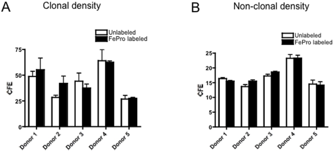
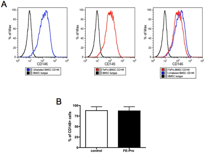
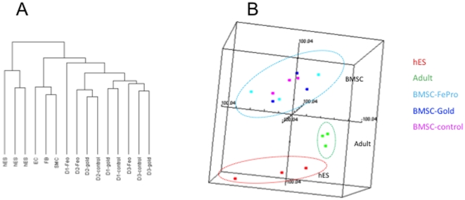
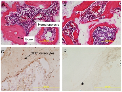
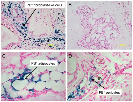
Superparamagnetic iron oxide nanoparticles (SPION) are increasingly used to label human bone marrow stromal cells (BMSCs, also called "mesenchymal stem cells") to monitor their fate by in vivo MRI, and by histology after Prussian blue (PB) staining. SPION-labeling appears to be safe as assessed by in vitro differentiation of BMSCs, however, we chose to resolve the question of the effect of labeling on maintaining the "stemness" of cells within the BMSC population in vivo. Assays performed include colony forming efficiency, CD146 expression, gene expression profiling, and the "gold standard" of evaluating bone and myelosupportive stroma formation in vivo in immuncompromised recipients. SPION-labeling did not alter these assays. Comparable abundant bone with adjoining host hematopoietic cells were seen in cohorts of mice that were implanted with SPION-labeled or unlabeled BMSCs. PB+ adipocytes were noted, demonstrating their donor origin, as well as PB+ pericytes, indicative of self-renewal of the stem cell in the BMSC population. This study confirms that SPION labeling does not alter the differentiation potential of the subset of stem cells within BMSCs.

摘要

超顺磁氧化铁纳米颗粒(SPION)越来越多地被用于标记人骨髓基质细胞(BMSCs,也称为“间充质干细胞”),通过体内 MRI 以及普鲁士蓝(PB)染色后的组织学来监测其命运。通过体外 BMSCs 分化评估,SPION 标记似乎是安全的,然而,我们选择解决标记对维持 BMSC 群体中细胞“干性”的体内影响的问题。进行的测定包括集落形成效率、CD146 表达、基因表达谱分析,以及在免疫缺陷受者体内评估骨和骨髓支持基质形成的“金标准”。SPION 标记并未改变这些测定。在植入 SPION 标记或未标记的 BMSCs 的小鼠队列中,观察到具有毗邻宿主造血细胞的可比丰富的骨。注意到 PB+脂肪细胞,表明其供体来源,以及 PB+周细胞,提示 BMSC 群体中的干细胞自我更新。这项研究证实,SPION 标记不会改变 BMSCs 中干细胞亚群的分化潜能。

https://cdn.ncbi.nlm.nih.gov/pmc/blobs/a2a9/2898800/1eacc6aa8e7b/pone.0011462.g006.jpg
https://cdn.ncbi.nlm.nih.gov/pmc/blobs/a2a9/2898800/1c0aa6e5f2a4/pone.0011462.g001.jpg
https://cdn.ncbi.nlm.nih.gov/pmc/blobs/a2a9/2898800/8235add8f62b/pone.0011462.g002.jpg
https://cdn.ncbi.nlm.nih.gov/pmc/blobs/a2a9/2898800/688d1d74810a/pone.0011462.g003.jpg
https://cdn.ncbi.nlm.nih.gov/pmc/blobs/a2a9/2898800/a0661b4a658a/pone.0011462.g004.jpg
https://cdn.ncbi.nlm.nih.gov/pmc/blobs/a2a9/2898800/1cacbf48570a/pone.0011462.g005.jpg
https://cdn.ncbi.nlm.nih.gov/pmc/blobs/a2a9/2898800/1eacc6aa8e7b/pone.0011462.g006.jpg

相似文献

1
Superparamagnetic iron oxide nanoparticles labeling of bone marrow stromal (mesenchymal) cells does not affect their "stemness".超顺磁性氧化铁纳米颗粒标记骨髓基质(间充质)细胞不会影响其“干性”。
PLoS One. 2010 Jul 7;5(7):e11462. doi: 10.1371/journal.pone.0011462.
2
In vivo tracing of superparamagnetic iron oxide-labeled bone marrow mesenchymal stem cells transplanted for traumatic brain injury by susceptibility weighted imaging in a rat model.在大鼠模型中,通过磁敏感加权成像对移植用于创伤性脑损伤的超顺磁性氧化铁标记的骨髓间充质干细胞进行体内示踪。
Chin J Traumatol. 2010 Jun 1;13(3):173-7.
3
Highly efficient magnetic stem cell labeling with citrate-coated superparamagnetic iron oxide nanoparticles for MRI tracking.高柠檬酸根修饰的超顺磁性氧化铁纳米颗粒高效标记干细胞及其磁共振示踪研究
Biomaterials. 2012 Jun;33(18):4515-25. doi: 10.1016/j.biomaterials.2012.02.064. Epub 2012 Mar 24.
4
Superparamagnetic iron oxide nanoparticles for direct labeling of stem cells and in vivo MRI tracking.用于干细胞直接标记和体内磁共振成像追踪的超顺磁性氧化铁纳米颗粒
Contrast Media Mol Imaging. 2016 Jan-Feb;11(1):55-64. doi: 10.1002/cmmi.1658. Epub 2015 Aug 3.
5
In vivo tracking of mesenchymal stem cells labeled with a novel chitosan-coated superparamagnetic iron oxide nanoparticles using 3.0T MRI.使用 3.0T MRI 对新型壳聚糖包被超顺磁性氧化铁纳米粒子标记的间充质干细胞进行体内示踪。
J Korean Med Sci. 2010 Feb;25(2):211-9. doi: 10.3346/jkms.2010.25.2.211. Epub 2010 Jan 21.
6
[In vivo tracking of bone marrow mesenchymal stem cells labeled with superparamagnetic iron oxide after cerebral ischemia in rats by magnetic resonance imaging].[磁共振成像对大鼠脑缺血后超顺磁性氧化铁标记的骨髓间充质干细胞的体内追踪]
Zhongguo Yi Xue Ke Xue Yuan Xue Bao. 2007 Feb;29(1):73-7.
7
MRI Tracking of SPIO- and -Labeled Bone Marrow Mesenchymal Stromal Cell Transplantation for Treatment of Stroke.MRI 示踪超顺磁性氧化铁标记骨髓间充质干细胞移植治疗脑卒中
Contrast Media Mol Imaging. 2019 Aug 29;2019:5184105. doi: 10.1155/2019/5184105. eCollection 2019.
8
MRI tracking of bone marrow mesenchymal stem cells labeled with ultra-small superparamagnetic iron oxide nanoparticles in a rat model of temporal lobe epilepsy.超小型超顺磁性氧化铁纳米颗粒标记的骨髓间充质干细胞在颞叶癫痫大鼠模型中的MRI追踪
Neurosci Lett. 2015 Oct 8;606:30-5. doi: 10.1016/j.neulet.2015.08.040. Epub 2015 Aug 25.
9
MR tracking of magnetically labeled mesenchymal stem cells in rats with liver fibrosis.磁共振示踪磁标记骨髓间充质干细胞在肝纤维化大鼠中的研究。
Magn Reson Imaging. 2010 Apr;28(3):394-9. doi: 10.1016/j.mri.2009.12.005. Epub 2010 Jan 21.
10
Labeling of cells with ferumoxides-protamine sulfate complexes does not inhibit function or differentiation capacity of hematopoietic or mesenchymal stem cells.用硫酸鱼精蛋白-铁氧化物复合物标记细胞不会抑制造血干细胞或间充质干细胞的功能或分化能力。
NMR Biomed. 2005 Dec;18(8):553-9. doi: 10.1002/nbm.991.

引用本文的文献

1
Magnetic Cell Targeting for Cardiovascular Tissue Engineering.用于心血管组织工程的磁性细胞靶向
Tissue Eng Part B Rev. 2025 Jun;31(3):234-247. doi: 10.1089/ten.TEB.2024.0103. Epub 2024 Aug 19.
2
Self-Organization of Iron Sulfide Nanoparticles into Complex Multicompartment Supraparticles.硫化亚铁纳米颗粒自组装成复杂的多隔室超粒子。
Adv Mater. 2023 Jun;35(23):e2211244. doi: 10.1002/adma.202211244. Epub 2023 Apr 25.
3
Controlling water-mediated interactions by designing self-assembled monolayer coatings.通过设计自组装单分子层涂层来控制水介导的相互作用。

本文引用的文献

1
Global transcriptional analysis for biomarker discovery and validation in cellular therapies.用于细胞疗法中生物标志物发现与验证的全转录组分析。
Mol Diagn Ther. 2009;13(3):181-93. doi: 10.2165/01250444-200913030-00003.
2
Microgel iron oxide nanoparticles for tracking human fetal mesenchymal stem cells through magnetic resonance imaging.用于通过磁共振成像追踪人胎儿间充质干细胞的微凝胶氧化铁纳米颗粒。
Stem Cells. 2009 Aug;27(8):1921-31. doi: 10.1002/stem.112.
3
Mesenchymal stem cells: revisiting history, concepts, and assays.间充质干细胞:重温历史、概念与检测方法
Sci Rep. 2021 Apr 19;11(1):8459. doi: 10.1038/s41598-021-87708-8.
4
Options for imaging cellular therapeutics in vivo: a multi-stakeholder perspective.体内细胞治疗成像选择:多利益相关者视角。
Cytotherapy. 2021 Sep;23(9):757-773. doi: 10.1016/j.jcyt.2021.02.005. Epub 2021 Apr 6.
5
Experimental and mathematical modelling of magnetically labelled mesenchymal stromal cell delivery.磁标记间充质基质细胞递送的实验和数学建模。
J R Soc Interface. 2021 Feb;18(175):20200558. doi: 10.1098/rsif.2020.0558. Epub 2021 Feb 17.
6
Superparamagnetic Iron Oxide Particles (VSOPs) Show Genotoxic Effects but No Functional Impact on Human Adipose Tissue-Derived Stromal Cells (ASCs).超顺磁性氧化铁颗粒(VSOPs)显示出遗传毒性作用,但对人脂肪组织来源的基质细胞(ASCs)无功能影响。
Materials (Basel). 2021 Jan 7;14(2):263. doi: 10.3390/ma14020263.
7
Applications of superparamagnetic iron oxide nanoparticles in drug and therapeutic delivery, and biotechnological advancements.超顺磁性氧化铁纳米颗粒在药物与治疗递送以及生物技术进展中的应用。
Beilstein J Nanotechnol. 2020 Jul 27;11:1092-1109. doi: 10.3762/bjnano.11.94. eCollection 2020.
8
Toxicity and Functional Impairment in Human Adipose Tissue-Derived Stromal Cells (hASCs) Following Long-Term Exposure to Very Small Iron Oxide Particles (VSOPs).长期暴露于超小氧化铁颗粒(VSOPs)后人脂肪组织来源的间充质干细胞(hASCs)的毒性和功能损伤
Nanomaterials (Basel). 2020 Apr 13;10(4):741. doi: 10.3390/nano10040741.
9
Use of nanoparticles in skeletal tissue regeneration and engineering.纳米粒子在骨骼组织再生和工程中的应用。
Histol Histopathol. 2020 Apr;35(4):331-350. doi: 10.14670/HH-18-184. Epub 2019 Nov 13.
10
Magnetically Assisted Control of Stem Cells Applied in 2D, 3D and In Situ Models of Cell Migration.磁场辅助控制干细胞在二维、三维和细胞迁移原位模型中的应用。
Molecules. 2019 Apr 19;24(8):1563. doi: 10.3390/molecules24081563.
Cell Stem Cell. 2008 Apr 10;2(4):313-9. doi: 10.1016/j.stem.2008.03.002.
4
Self-renewing osteoprogenitors in bone marrow sinusoids can organize a hematopoietic microenvironment.骨髓血窦中的自我更新骨祖细胞能够构建造血微环境。
Cell. 2007 Oct 19;131(2):324-36. doi: 10.1016/j.cell.2007.08.025.
5
Postnatal skeletal stem cells.产后骨骼干细胞
Methods Enzymol. 2006;419:117-48. doi: 10.1016/S0076-6879(06)19006-0.
6
Tracking neural stem cells in patients with brain trauma.追踪脑外伤患者体内的神经干细胞。
N Engl J Med. 2006 Nov 30;355(22):2376-8. doi: 10.1056/NEJMc055304.
7
Expression of transferrin receptor and ferritin following ferumoxides-protamine sulfate labeling of cells: implications for cellular magnetic resonance imaging.硫酸鱼精蛋白-超顺磁性氧化铁标记细胞后转铁蛋白受体和铁蛋白的表达:对细胞磁共振成像的意义
NMR Biomed. 2006 Aug;19(5):581-92. doi: 10.1002/nbm.1038.
8
Magnetic resonance tracking of dendritic cells in melanoma patients for monitoring of cellular therapy.黑色素瘤患者中树突状细胞的磁共振追踪用于细胞治疗监测
Nat Biotechnol. 2005 Nov;23(11):1407-13. doi: 10.1038/nbt1154. Epub 2005 Oct 30.
9
Labeling of cells with ferumoxides-protamine sulfate complexes does not inhibit function or differentiation capacity of hematopoietic or mesenchymal stem cells.用硫酸鱼精蛋白-铁氧化物复合物标记细胞不会抑制造血干细胞或间充质干细胞的功能或分化能力。
NMR Biomed. 2005 Dec;18(8):553-9. doi: 10.1002/nbm.991.
10
Magnetic resonance imaging and confocal microscopy studies of magnetically labeled endothelial progenitor cells trafficking to sites of tumor angiogenesis.磁标记内皮祖细胞向肿瘤血管生成部位迁移的磁共振成像和共聚焦显微镜研究。
Stem Cells. 2006 Mar;24(3):671-8. doi: 10.1634/stemcells.2005-0017. Epub 2005 Sep 22.