Cardiology Division, Department of Medicine, Gazes Cardiac Research Institute, Medical University of South Carolina, Charleston, South Carolina, United States of America.
PLoS One. 2010 Jul 12;5(7):e11470. doi: 10.1371/journal.pone.0011470.
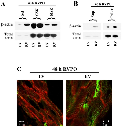
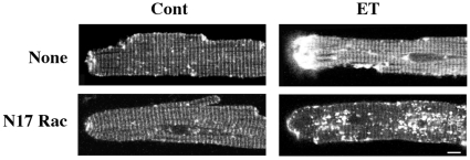
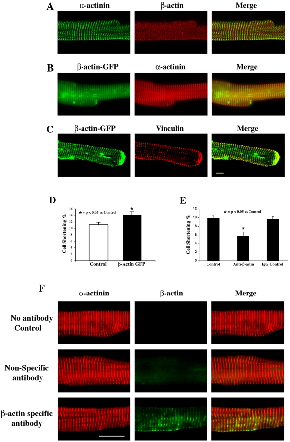
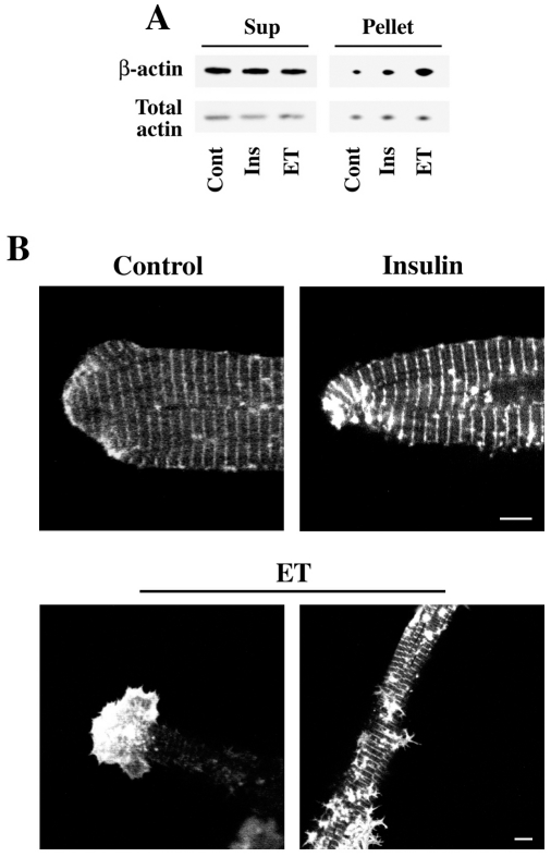
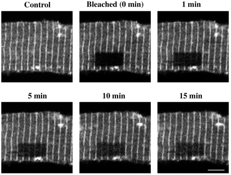
The myocardium responds to hemodynamic stress through cellular growth and organ hypertrophy. The impact of cytoskeletal elements on this process, however, is not fully understood. While alpha-actin in cardiomyocytes governs muscle contraction in combination with the myosin motor, the exact role of beta-actin has not been established. We hypothesized that in adult cardiomyocytes, as in non-myocytes, beta-actin can facilitate cytoskeletal rearrangement within cytoskeletal structures such as Z-discs. Using a feline right ventricular pressure overload (RVPO) model, we measured the level and distribution of beta-actin in normal and pressure overloaded myocardium. Resulting data demonstrated enriched levels of beta-actin and enhanced translocation to the Triton-insoluble cytoskeletal and membrane skeletal complexes. In addition, RVPO in vivo and in vitro hypertrophic stimulation with endothelin (ET) or insulin in isolated adult cardiomyocytes enhanced the content of polymerized fraction (F-actin) of beta-actin. To determine the localization and dynamics of beta-actin, we adenovirally expressed GFP-tagged beta-actin in isolated adult cardiomyocytes. The ectopically expressed beta-actin-GFP localized to the Z-discs, costameres, and cell termini. Fluorescence recovery after photobleaching (FRAP) measurements of beta-actin dynamics revealed that beta-actin at the Z-discs is constantly being exchanged with beta-actin from cytoplasmic pools and that this exchange is faster upon hypertrophic stimulation with ET or insulin. In addition, in electrically stimulated isolated adult cardiomyocytes, while beta-actin overexpression improved cardiomyocyte contractility, immunoneutralization of beta-actin resulted in a reduced contractility suggesting that beta-actin could be important for the contractile function of adult cardiomyocytes. These studies demonstrate the presence and dynamics of beta-actin in the adult cardiomyocyte and reinforce its usefulness in measuring cardiac cytoskeletal rearrangement during hypertrophic stimulation.
心肌通过细胞生长和器官肥大来应对血液动力应激。然而,细胞骨架元素对此过程的影响尚不完全清楚。虽然心肌细胞中的α-肌动蛋白与肌球蛋白马达一起控制肌肉收缩,但β-肌动蛋白的确切作用尚未确定。我们假设,在成年心肌细胞中,与非心肌细胞一样,β-肌动蛋白可以促进细胞骨架结构(如 Z 盘)内的细胞骨架重排。我们使用猫右心室压力超负荷(RVPO)模型,测量了正常和压力超负荷心肌中β-肌动蛋白的水平和分布。结果数据表明,β-肌动蛋白水平丰富,并向 Triton 不溶性细胞骨架和膜骨架复合物易位增强。此外,体内 RVPO 和体外用内皮素(ET)或胰岛素刺激分离的成年心肌细胞,增强了β-肌动蛋白聚合部分(F-肌动蛋白)的含量。为了确定β-肌动蛋白的定位和动态,我们在分离的成年心肌细胞中用腺病毒表达 GFP 标记的β-肌动蛋白。异位表达的β-肌动蛋白-GFP 定位于 Z 盘、肌节和细胞末端。β-肌动蛋白动力学的荧光恢复后光漂白(FRAP)测量表明,Z 盘上的β-肌动蛋白与细胞质池中的β-肌动蛋白不断交换,而 ET 或胰岛素刺激引起的肥大时,这种交换更快。此外,在电刺激分离的成年心肌细胞中,虽然β-肌动蛋白过表达可改善心肌细胞的收缩性,但β-肌动蛋白的免疫中和导致收缩性降低,这表明β-肌动蛋白对成年心肌细胞的收缩功能可能很重要。这些研究表明β-肌动蛋白在成年心肌细胞中的存在和动态,并强调了其在测量肥大刺激期间心脏细胞骨架重排中的有用性。