Department of Diagnostic and Interventional Radiology, University Hospital Tuebingen, Hoppe-Seyler-Strasse 3, Tuebingen, Germany.
Korean J Radiol. 2010 Sep-Oct;11(5):547-52. doi: 10.3348/kjr.2010.11.5.547. Epub 2010 Aug 27.
We aimed to estimate the effective dose of 4D-Perfusion-CT protocols of the lung, liver, and pelvis for the assessment of tumor vascularity.
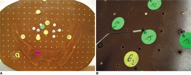
An Alderson-Rando phantom equipped with thermoluminescent dosimeters was used to determine the effective dose values of 4D-Perfusion-CT. Phantom measurements were performed on a 128-slice single-source scanner in adaptive 4D-spiral-mode with bidirectional table movement and a total scan range of 69 mm over a time period of nearly 120 seconds (26 scans). Perfusion measurements were simulated for the lung, liver, and pelvis under the following conditions: lung (80 kV, 60 mAs), liver (80 kV/80 mAs and 80 kV/120 mAs), pelvis (100 kV/80 mAs and 100 kV/120 mAs).
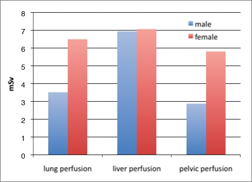
Depending on gender, the evaluated body region and scan protocol, an effective whole-body dose between 2.9-12.2 mSv, was determined. The radiation exposure administered to gender-specific organs like the female breast tissue (lung perfusion) or to the ovaries (pelvic perfusion) led to an increase in the female specific dose by 86% and 100% in perfusion scans of the lung and the pelvis, respectively.
Due to a significant radiation dose of 4D-perfusion-CT protocols, the responsible use of this new promising technique is mandatory. Gender- and organ-specific differences should be considered for indication and planning of tumor perfusion scans.
我们旨在评估肺部、肝脏和骨盆的 4D 灌注 CT 方案评估肿瘤血管生成的有效剂量。
使用配备有热释光剂量计的 Alderson-Rando 体模来确定 4D 灌注 CT 的有效剂量值。在自适应 4D 螺旋模式下,使用 128 层单源扫描仪对体模进行了测量,该模式采用双向台移动,总扫描范围为近 120 秒(26 次扫描)的 69 毫米。在以下条件下模拟了肺部、肝脏和骨盆的灌注测量:肺部(80 kV,60 mAs)、肝脏(80 kV/80 mAs 和 80 kV/120 mAs)、骨盆(100 kV/80 mAs 和 100 kV/120 mAs)。
根据性别、评估的身体区域和扫描方案,确定了全身有效剂量在 2.9-12.2 mSv 之间。对女性乳房组织(肺部灌注)或卵巢(骨盆灌注)等特定性别器官进行的辐射暴露导致肺部和骨盆灌注扫描的女性特定剂量分别增加了 86%和 100%。
由于 4D 灌注 CT 方案的辐射剂量显著,因此必须负责任地使用这项新的有前途的技术。在指示和规划肿瘤灌注扫描时,应考虑性别和器官特异性差异。