Laboratory of Electron Microscopy Pietro M. Motta, Department of Human Anatomic, Histologic, Forensic and Locomotor Apparatus Sciences, Sapienza University of Rome, via Alfonso Borelli 50, Rome, Italy.
Eur J Histochem. 2010 Jul 14;54(3):e33. doi: 10.4081/ejh.2010.e33.
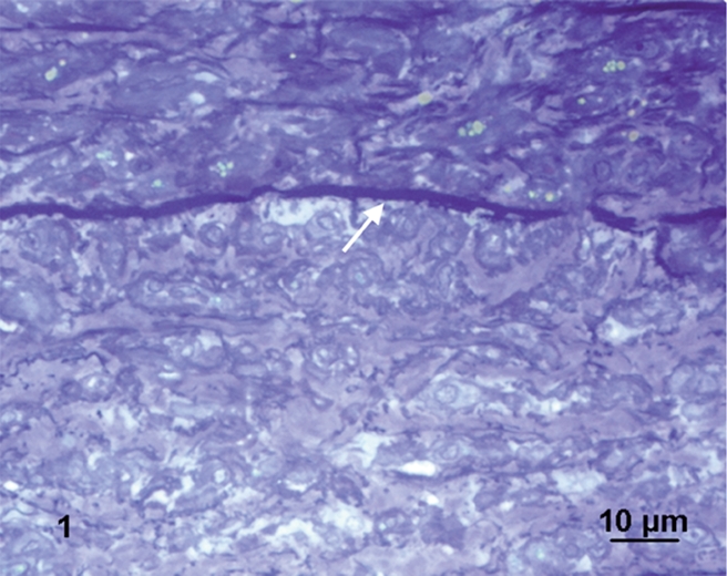
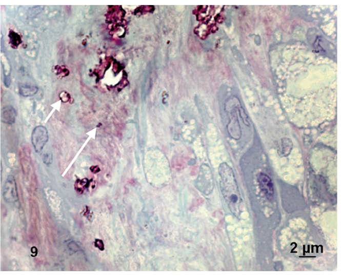
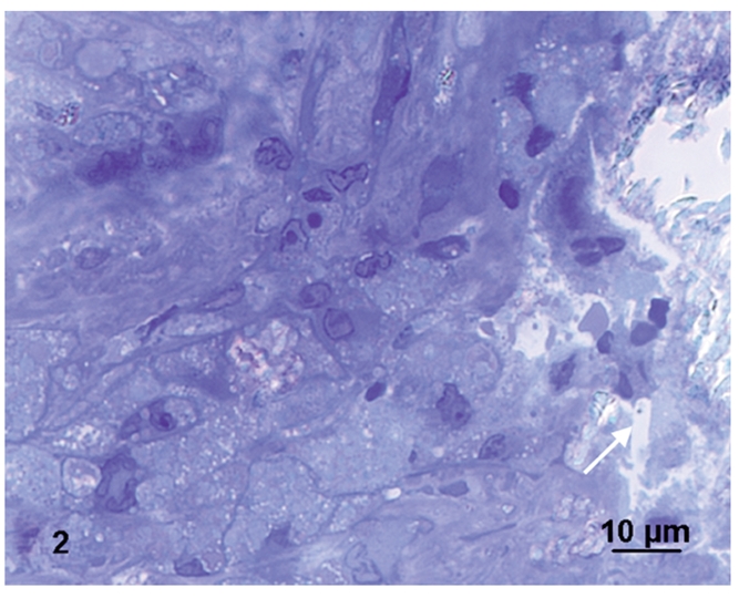
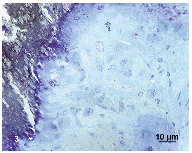
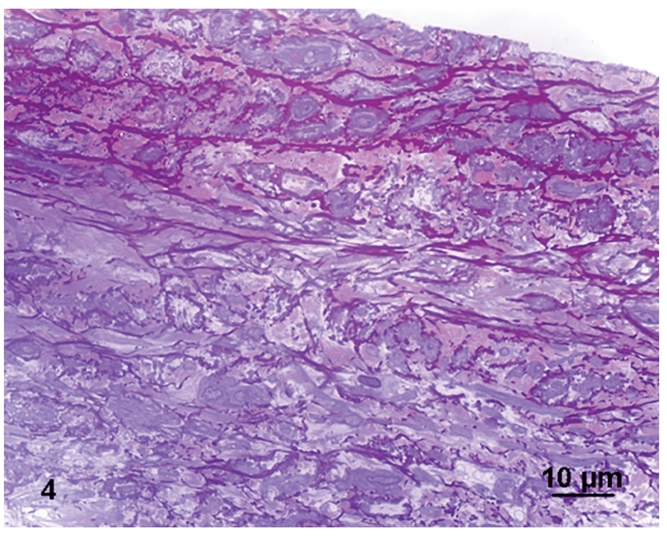
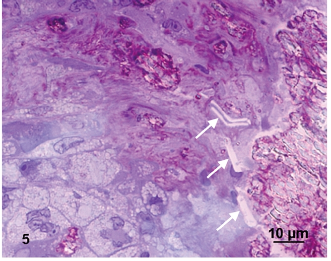
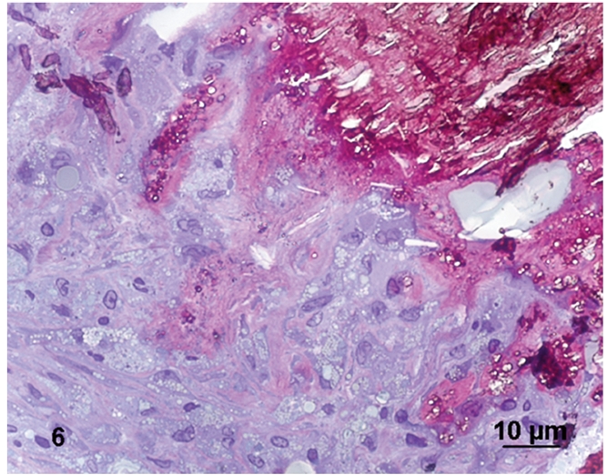
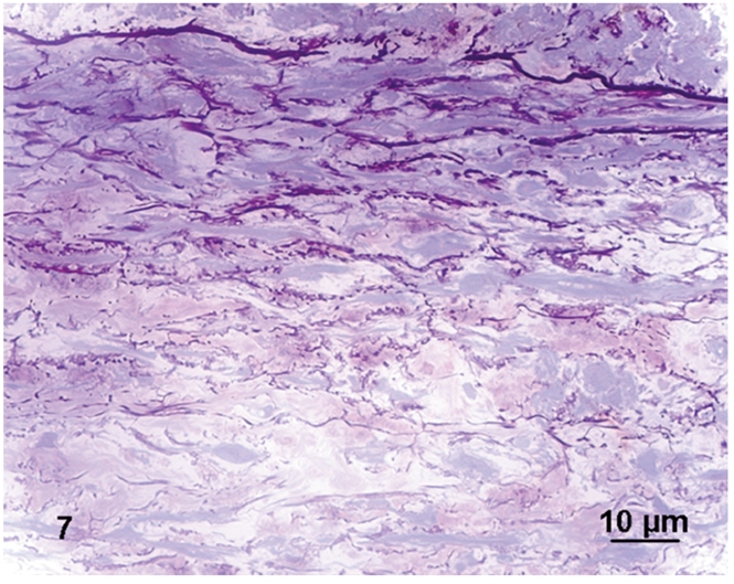
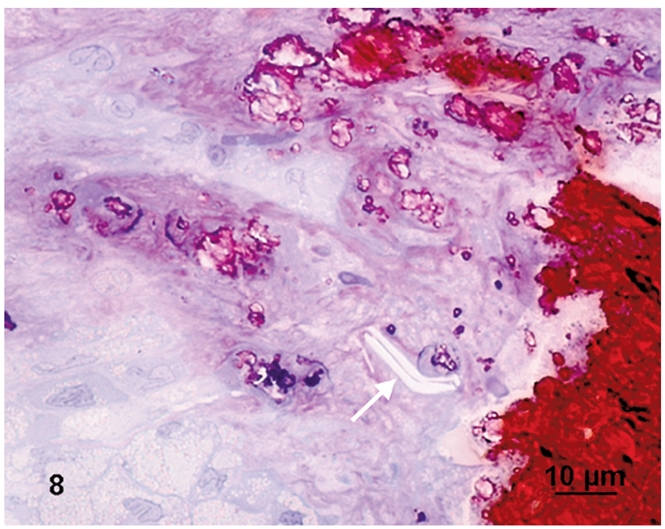
Atherosclerotic plaques have a high probability of undergoing rapid progression to stenosis, becoming responsible of acute coronary syndrome or stroke. Microcalcifications may act as enhancers of atherosclerotic plaque vulnerability. Considering that calcifications with a diameter smaller than 10 mm in paraffin embedded tissue are rather difficult to detect, our aim was to analyze microcalcifications on semithin sections from epoxy resin embedded samples of carotid endarterectomies using an original trichromic stain (methylene blue--azur B--basic fuchsine--alizarin red). We have compared samples stained either with our method, methylene blue-azur B alone or with Von Kossa staining, and methylene blue-azur B -basic fuchsine alone or with Von Kossa staining. Our method resulted to be simple and fast (ca. 2 min), it gives a sharp general contrast for all structures and allows to easy identify collagen and elastin. In addition, gray-green colour associated to intracellular lipid droplets evidences foam cells, which are particularly abundant in endarterectomies samples. Mast cells and their metachromatic granules are also well recognized. Calcifications over 0,5 mm are clearly recognizable. In conclusion, microcalcifications are clearly distinguished from the extracellular matrix in spite of their reduced dimensions. Methylene blue--azur B--basic fuchsine--alizarin red method is easy to use, reproducible, and is particularly suitable for the identification of microcalcifications in the morphological analysis of atherosclerotic plaques.
动脉粥样硬化斑块极有可能迅速进展为狭窄,导致急性冠脉综合征或中风。微钙化可能会增强动脉粥样硬化斑块的脆弱性。考虑到石蜡包埋组织中直径小于 10mm 的钙化较难检测到,我们的目的是使用一种原始的三色染色法(亚甲蓝-天青 B-碱性品红-茜素红)分析颈动脉内膜切除术环氧包埋样本的半薄切片上的微钙化。我们比较了用我们的方法、亚甲蓝-天青 B 单独或与 Von Kossa 染色、亚甲蓝-天青 B-碱性品红单独或与 Von Kossa 染色染色的样本。我们的方法简单快捷(约 2 分钟),可以为所有结构提供鲜明的整体对比度,并易于识别胶原和弹性蛋白。此外,与细胞内脂质滴相关的灰绿色颜色表明泡沫细胞,泡沫细胞在内膜切除术样本中特别丰富。肥大细胞及其异染颗粒也能很好地识别。超过 0.5mm 的钙化可以清晰识别。总之,尽管微钙化的尺寸较小,但与细胞外基质仍能清晰区分。亚甲蓝-天青 B-碱性品红-茜素红方法易于使用、可重复,特别适用于动脉粥样硬化斑块形态分析中微钙化的鉴定。