Periodontics Division, University of Brasilia, Brasília, DF, Brazil.
J Appl Oral Sci. 2010 Jul-Aug;18(4):379-84. doi: 10.1590/s1678-77572010000400010.
This study assessed the bone density gain and its relationship with the periodontal clinical parameters in a case series of a regenerative therapy procedure.
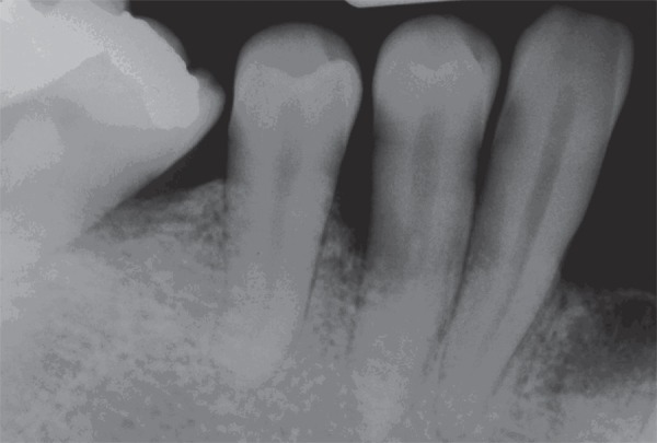
Using a split-mouth study design, 10 pairs of infrabony defects from 15 patients were treated with a pool of bovine bone morphogenetic proteins associated with collagen membrane (test sites) or collagen membrane only (control sites). The periodontal healing was clinically and radiographically monitored for six months. Standardized pre-surgical and 6-month postoperative radiographs were digitized for digital subtraction analysis, which showed relative bone density gain in both groups of 0.034 ± 0.423 and 0.105 ± 0.423 in the test and control group, respectively (p>0.05).
As regards the area size of bone density change, the influence of the therapy was detected in 2.5 mm² in the test group and 2 mm² in the control group (p>0.05). Additionally, no correlation was observed between the favorable clinical results and the bone density gain measured by digital subtraction radiography (p>0.05).
The findings of this study suggest that the clinical benefit of the regenerative therapy observed did not come with significant bone density gains. Long-term evaluation may lead to a different conclusions.
本研究通过一系列再生治疗病例评估了骨密度的增加及其与牙周临床参数的关系。
采用分牙区对照研究设计,15 名患者的 10 对骨下缺损分别接受牛骨形态发生蛋白联合胶原膜(实验组)或仅胶原膜(对照组)治疗。对牙周愈合情况进行了 6 个月的临床和放射学监测。对术前和术后 6 个月的标准放射照片进行数字化,进行数字减影分析,结果显示实验组和对照组的相对骨密度分别增加了 0.034±0.423 和 0.105±0.423(p>0.05)。
在骨密度变化的面积大小方面,实验组的治疗效果为 2.5mm²,对照组为 2mm²(p>0.05)。此外,数字减影放射摄影测量的骨密度增加与临床疗效之间未观察到相关性(p>0.05)。
本研究结果表明,观察到的再生治疗的临床获益并未带来显著的骨密度增加。长期评估可能会得出不同的结论。