• 文献检索
  • 文档翻译
  • 深度研究
  • 学术资讯
  • Suppr Zotero 插件Zotero 插件
  • 邀请有礼
  • 套餐&价格
  • 历史记录
应用&插件
Suppr Zotero 插件Zotero 插件浏览器插件Mac 客户端Windows 客户端微信小程序
定价
高级版会员购买积分包购买API积分包
服务
文献检索文档翻译深度研究API 文档MCP 服务
关于我们
关于 Suppr公司介绍联系我们用户协议隐私条款
关注我们

Suppr 超能文献

核心技术专利:CN118964589B侵权必究
粤ICP备2023148730 号-1Suppr @ 2026

文献检索

告别复杂PubMed语法,用中文像聊天一样搜索,搜遍4000万医学文献。AI智能推荐,让科研检索更轻松。

立即免费搜索

文件翻译

保留排版,准确专业,支持PDF/Word/PPT等文件格式,支持 12+语言互译。

免费翻译文档

深度研究

AI帮你快速写综述,25分钟生成高质量综述,智能提取关键信息,辅助科研写作。

立即免费体验

相似文献

1
In vivo magnetic resonance imaging and optical imaging comparison of viable and nonviable mesenchymal stem cells with a bifunctional label.活细胞与死细胞间充质干细胞的双功能标记物的体内磁共振成像和光学成像比较。
Mol Imaging. 2010 Oct;9(5):278-90.
2
[In vivo MR imaging tracking of supermagnetic iron-oxide nanoparticle-labeled bone marrow mesenchymal stem cells injected into intra-articular space of knee joints: experiment with rabbit].超顺磁性氧化铁纳米颗粒标记的骨髓间充质干细胞注入膝关节腔的体内磁共振成像追踪:兔实验
Zhonghua Yi Xue Za Zhi. 2007 Dec 4;87(45):3213-8.
3
Magnetic resonance imaging of ferumoxide-labeled mesenchymal stem cells in cartilage defects: in vitro and in vivo investigations.铁氧化物标记的间充质干细胞在软骨缺损中的磁共振成像:体外和体内研究。
Mol Imaging. 2012 Jun;11(3):197-209.
4
In vivo MR imaging tracking of magnetic iron oxide nanoparticle labeled, engineered, autologous bone marrow mesenchymal stem cells following intra-articular injection.关节内注射磁性氧化铁纳米颗粒标记的、经工程改造的自体骨髓间充质干细胞后的体内磁共振成像跟踪
Joint Bone Spine. 2008 Jul;75(4):432-8. doi: 10.1016/j.jbspin.2007.09.013. Epub 2008 May 2.
5
Transplantation of Aggregates of Autologous Synovial Mesenchymal Stem Cells for Treatment of Cartilage Defects in the Femoral Condyle and the Femoral Groove in Microminipigs.自体滑膜间充质干细胞聚集物移植治疗小型猪股骨髁和股骨沟软骨缺损。
Am J Sports Med. 2019 Aug;47(10):2338-2347. doi: 10.1177/0363546519859855. Epub 2019 Jul 15.
6
Magnetic resonance imaging of mesenchymal stem cells labeled with dual (MR and fluorescence) agents in rat spinal cord injury.大鼠脊髓损伤中用双(磁共振和荧光)标记剂标记的间充质干细胞的磁共振成像
Acad Radiol. 2009 Sep;16(9):1142-54. doi: 10.1016/j.acra.2009.03.016.
7
Trimodal Cell Tracking In Vivo: Combining Iron- and Fluorine-Based Magnetic Resonance Imaging with Magnetic Particle Imaging to Monitor the Delivery of Mesenchymal Stem Cells and the Ensuing Inflammation.体内三模态细胞追踪:结合基于铁和氟的磁共振成像与磁性粒子成像以监测间充质干细胞的递送及随后的炎症反应。
Tomography. 2019 Dec;5(4):367-376. doi: 10.18383/j.tom.2019.00020.
8
MR tracking of magnetically labeled mesenchymal stem cells in rat kidneys with acute renal failure.急性肾衰竭大鼠肾脏中磁性标记间充质干细胞的磁共振追踪
Cell Transplant. 2008;17(3):279-90. doi: 10.3727/096368908784153878.
9
In vivo tracking and fate of intra-articularly injected superparamagnetic iron oxide particle-labeled multipotent stromal cells in an ovine model of osteoarthritis.在骨关节炎绵羊模型中,关节内注射超顺磁性氧化铁颗粒标记的多能间充质干细胞的体内追踪及转归
Cell Transplant. 2015;24(11):2379-90. doi: 10.3727/096368914X685654. Epub 2014 Dec 12.
10
In vivo quantitative assessment of cell viability of gadolinium or iron-labeled cells using MRI and bioluminescence imaging.采用 MRI 和生物发光成像技术对钆或铁标记细胞的体内细胞活力进行定量评估。
Contrast Media Mol Imaging. 2013 Mar-Apr;8(2):165-74. doi: 10.1002/cmmi.1513.

引用本文的文献

1
Clinical Tracking of Cell Transfer and Cell Transplantation: Trials and Tribulations.细胞转移和细胞移植的临床追踪:试验和磨难。
Radiology. 2018 Dec;289(3):604-615. doi: 10.1148/radiol.2018180449. Epub 2018 Oct 9.
2
Neural Differentiation of Bone Marrow Mesenchymal Stem Cells Carrying the FTH1 Reporter Gene and Detection with MRI.携带有 FTH1 报告基因的骨髓间充质干细胞的神经分化及其 MRI 检测。
Biomed Res Int. 2018 Jun 26;2018:1978602. doi: 10.1155/2018/1978602. eCollection 2018.
3
The Protein Corona around Nanoparticles Facilitates Stem Cell Labeling for Clinical MR Imaging.纳米颗粒周围的蛋白质冠层有助于用于临床磁共振成像的干细胞标记。
Radiology. 2018 Mar;286(3):938-947. doi: 10.1148/radiol.2017170130. Epub 2017 Nov 1.
4
Application prospective of nanoprobes with MRI and FI dual-modality imaging on breast cancer stem cells in tumor.具有MRI和FI双模态成像的纳米探针在肿瘤中乳腺癌干细胞的应用前景
J Nanobiotechnology. 2016 Jun 23;14(1):52. doi: 10.1186/s12951-016-0195-8.
5
Macrophage phagocytosis alters the MRI signal of ferumoxytol-labeled mesenchymal stromal cells in cartilage defects.巨噬细胞吞噬作用改变了软骨缺损中铁氧还蛋白标记的间充质基质细胞的MRI信号。
Sci Rep. 2016 May 13;6:25897. doi: 10.1038/srep25897.
6
Clinically applicable magnetic-labeling of natural killer cells for MRI of transcatheter delivery to liver tumors: preclinical validation for clinical translation.用于经导管递送至肝肿瘤的MRI的自然杀伤细胞的临床适用磁性标记:临床转化的临床前验证
Nanomedicine (Lond). 2015;10(11):1761-74. doi: 10.2217/nnm.15.24.
7
Tracking fusion of human mesenchymal stem cells after transplantation to the heart.追踪人类间充质干细胞移植至心脏后的融合情况。
Stem Cells Transl Med. 2015 Jun;4(6):685-94. doi: 10.5966/sctm.2014-0198. Epub 2015 Apr 6.
8
Magnetic resonance imaging of stem cell apoptosis in arthritic joints with a caspase activatable contrast agent.使用半胱天冬酶激活造影剂对关节炎关节中干细胞凋亡进行磁共振成像。
ACS Nano. 2015 Feb 24;9(2):1150-60. doi: 10.1021/nn504494c. Epub 2015 Jan 26.
9
Molecular imaging, biodistribution and efficacy of mesenchymal bone marrow cell therapy in a mouse model of Chagas disease.查加斯病小鼠模型中间充质骨髓细胞治疗的分子成像、生物分布及疗效
Microbes Infect. 2014 Nov;16(11):923-935. doi: 10.1016/j.micinf.2014.08.016. Epub 2014 Sep 16.
10
MR Imaging of Stem Cell Transplants in Arthritic Joints.关节炎关节中干细胞移植的磁共振成像
J Stem Cell Res Ther. 2014 Feb 7;4(2):165. doi: 10.4172/2157-7633.1000165.

本文引用的文献

1
Labeling human mesenchymal stem cells with fluorescent contrast agents: the biological impact.用荧光对比剂标记人骨髓间充质干细胞:生物学影响。
Mol Imaging Biol. 2011 Feb;13(1):3-9. doi: 10.1007/s11307-010-0322-0.
2
An optical imaging method to monitor stem cell migration in a model of immune-mediated arthritis.一种用于监测免疫介导性关节炎模型中干细胞迁移的光学成像方法。
Opt Express. 2009 Dec 21;17(26):24403-13. doi: 10.1364/OE.17.024403.
3
The influence of ferucarbotran on the chondrogenesis of human mesenchymal stem cells.瑞必安对人间充质干细胞软骨形成的影响。
Contrast Media Mol Imaging. 2009 Jul-Aug;4(4):165-73. doi: 10.1002/cmmi.276.
4
Relaxation effects of ferucarbotran-labeled mesenchymal stem cells at 1.5T and 3T: discrimination of viable from lysed cells.阿魏酸瑞香素标记的间充质干细胞在1.5T和3T磁场下的弛豫效应:活细胞与裂解细胞的鉴别
Magn Reson Med. 2009 Aug;62(2):325-32. doi: 10.1002/mrm.22011.
5
Cell tracking with optical imaging.光学成像细胞追踪
Eur Radiol. 2008 Oct;18(10):2021-32. doi: 10.1007/s00330-008-0984-z. Epub 2008 May 28.
6
Delivery of fluorescent probes using iron oxide particles as carriers enables in-vivo labeling of migrating neural precursors for magnetic resonance imaging and optical imaging.使用氧化铁颗粒作为载体递送荧光探针能够对迁移的神经前体细胞进行体内标记,用于磁共振成像和光学成像。
J Biomed Opt. 2007 Sep-Oct;12(5):051504. doi: 10.1117/1.2800294.
7
Magneto-optical labeling of fetal neural stem cells for in vivo MRI tracking.用于体内MRI追踪的胎儿神经干细胞的磁光标记
Conf Proc IEEE Eng Med Biol Soc. 2006;2006:5631-4. doi: 10.1109/IEMBS.2006.259982.
8
Molecular imaging: integration of molecular imaging into the musculoskeletal imaging practice.分子成像:将分子成像融入肌肉骨骼成像实践
Radiology. 2007 Sep;244(3):651-71. doi: 10.1148/radiol.2443060295.
9
Cell tracking using magnetic resonance imaging.使用磁共振成像进行细胞追踪。
J Physiol. 2007 Oct 1;584(Pt 1):25-30. doi: 10.1113/jphysiol.2007.139451. Epub 2007 Aug 9.
10
Iron-oxide labeling of hematogenous macrophages in a model of experimental autoimmune encephalomyelitis and the contribution to signal loss in fast imaging employing steady state acquisition (FIESTA) images.实验性自身免疫性脑脊髓炎模型中血源性巨噬细胞的氧化铁标记及其对稳态采集快速成像(FIESTA)图像信号丢失的影响
J Magn Reson Imaging. 2007 Jul;26(1):144-51. doi: 10.1002/jmri.21005.

活细胞与死细胞间充质干细胞的双功能标记物的体内磁共振成像和光学成像比较。

In vivo magnetic resonance imaging and optical imaging comparison of viable and nonviable mesenchymal stem cells with a bifunctional label.

机构信息

Department of Radiology, Mount Auburn Hospital, 330 Mount Auburn Street, Cambridge, MA 02138, USA.

出版信息

Mol Imaging. 2010 Oct;9(5):278-90.

PMID:20868628
原文链接:https://pmc.ncbi.nlm.nih.gov/articles/PMC3060764/
Abstract

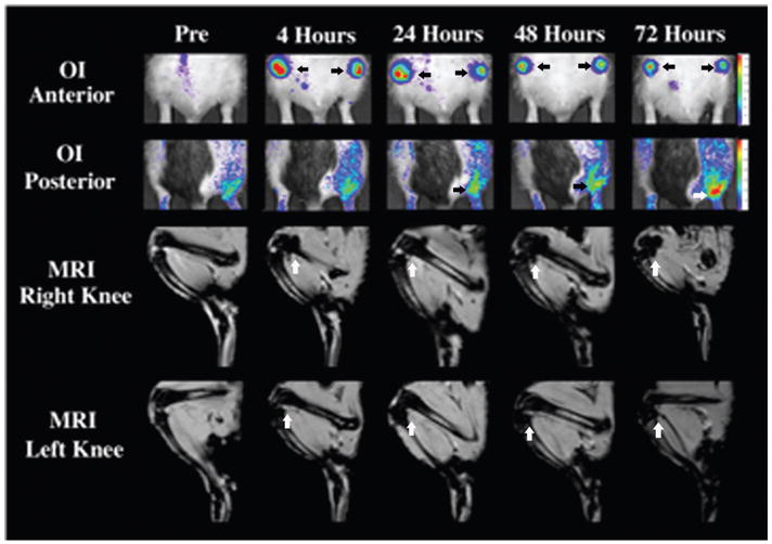
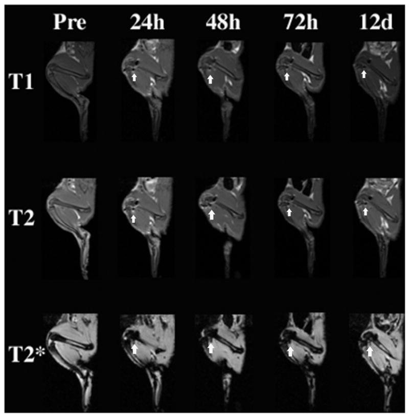
The purpose of this study was to compare viable and nonviable bilabeled mesenchymal stem cells (MSCs) in arthritic joints with magnetic resonance imaging (MRI) and optical imaging (OI). MSCs were labeled with ferucarbotran and DiD. MRI and OI of bilabeled cells were compared with controls. Six rats with arthritis received intra-articular injections of bilabeled viable MSCs into the right knee and nonviable MSCs into the left knee. Animals underwent MRI and OI preinjection and at 4, 24, 48, and 72 hours postinjection. The results were analyzed with a mixed random effects model and Fisher probability. Bilabeled MSCs showed increased MRI and OI signals compared to unlabeled controls (p < .0001). After intra-articular injection, bilabeled MSCs caused significant T2 and T2* effect on MRI and fluorescence on OI up to 72 hours postinjection (p < .05). There was no significant difference between viable and nonviable MSC signal in the knee joints; however, some of the viable cells migrated to an adjacent inflamed ankle joint (p < .05). Immunohistochemistry confirmed viable MSCs in right knee and ankle joints and nonviable MSCs in the left knee. Viable and nonviable cells could not be differentiated with MRI or OI signal intensity but were differentiated based on their ability to migrate in vivo.

摘要

本研究旨在通过磁共振成像(MRI)和光学成像(OI)比较关节炎关节中活细胞和死细胞双标记间充质干细胞(MSCs)。MSCs 用 Ferucarbotran 和 DiD 进行标记。将双标记活细胞和死细胞的 MRI 和 OI 与对照进行比较。6 只关节炎大鼠的右膝关节内注射双标记活 MSCs,左膝关节内注射非活 MSCs。动物在注射前、注射后 4、24、48 和 72 小时进行 MRI 和 OI。结果采用混合随机效应模型和 Fisher 概率进行分析。与未标记对照相比,双标记 MSCs 显示出增强的 MRI 和 OI 信号(p<0.0001)。关节内注射后,双标记 MSCs 在注射后 72 小时内对 MRI 产生显著的 T2 和 T2*效应,对 OI 产生荧光效应(p<0.05)。膝关节中活细胞和死细胞的信号无显著差异;然而,一些活细胞迁移到相邻的发炎踝关节(p<0.05)。免疫组织化学证实右膝关节和踝关节中有活的 MSCs,左膝关节中有非活的 MSCs。活细胞和死细胞不能通过 MRI 或 OI 信号强度来区分,但可以根据其在体内迁移的能力来区分。

https://cdn.ncbi.nlm.nih.gov/pmc/blobs/0e4f/3060764/69c7a15c67c0/nihms278548f6.jpg
https://cdn.ncbi.nlm.nih.gov/pmc/blobs/0e4f/3060764/886df47bbbd3/nihms278548f1.jpg
https://cdn.ncbi.nlm.nih.gov/pmc/blobs/0e4f/3060764/f84fa3bb3941/nihms278548f2.jpg
https://cdn.ncbi.nlm.nih.gov/pmc/blobs/0e4f/3060764/9e6f67e48451/nihms278548f3.jpg
https://cdn.ncbi.nlm.nih.gov/pmc/blobs/0e4f/3060764/09d3ac4a0532/nihms278548f4.jpg
https://cdn.ncbi.nlm.nih.gov/pmc/blobs/0e4f/3060764/be4737bf8834/nihms278548f5.jpg
https://cdn.ncbi.nlm.nih.gov/pmc/blobs/0e4f/3060764/69c7a15c67c0/nihms278548f6.jpg