Department of Academic Biochemistry, Institute of Cancer Research, London, UK.
Breast Cancer Res. 2010;12(5):R76. doi: 10.1186/bcr2719. Epub 2010 Sep 28.
Very few studies have investigated whether the time elapsed between surgical resection and tissue fixation or the difference between core-cut and excision biopsies impact on immunohistochemically measured biomarkers including phosphorylated proteins in primary breast cancer. The aim of this study was to characterize the differences in immunoreactivity of common biomarkers that may occur (a) due to tissue handling at surgery and (b) between core-cuts and resected tumours.
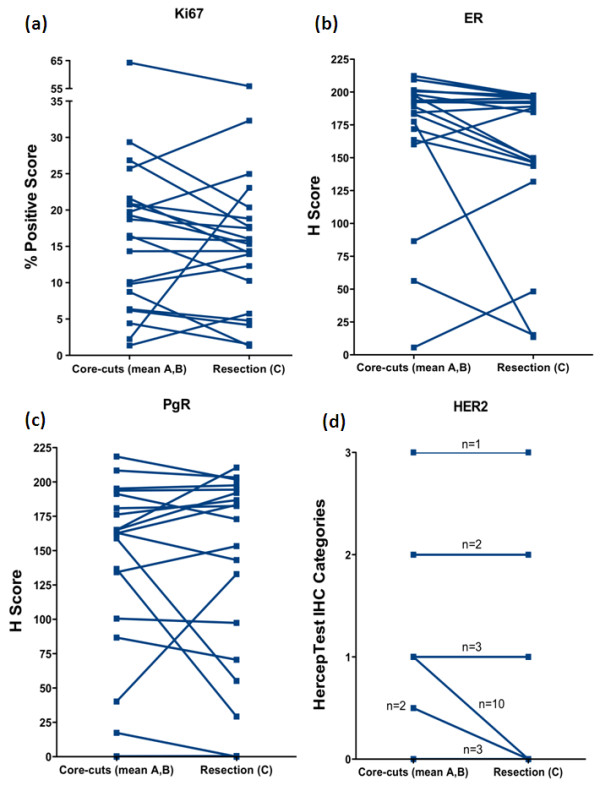
Core-cuts taken from surgical breast cancer specimens immediately after resection (sample A) and after routine X-ray of the excised tumour (sample B) were formalin-fixed and paraffin-embedded and compared to the routinely fixed resection specimen (sample C). The variation in immunohistochemical expression of Ki67, oestrogen receptor (ER), progesterone receptor (PgR), human epidermal growth factor 2 (HER2), p-Akt and p-Erk were investigated.
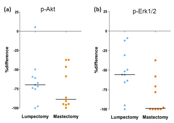
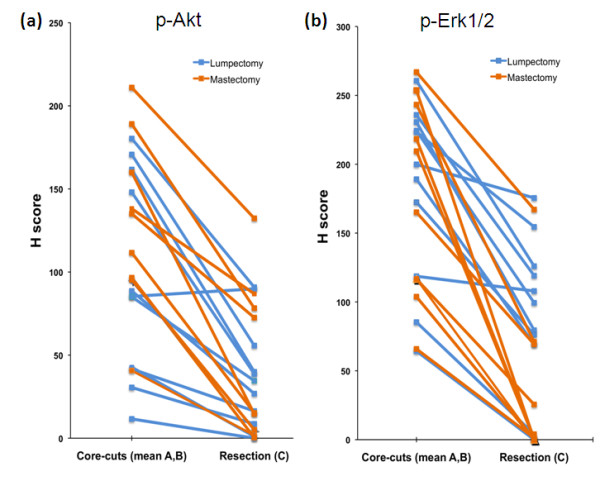
Twenty-one tissue sets with adequate tumour were available. Median time between collection of core-cuts A and B was 30 minutes (range 20 to 80). None of the markers showed significant differences between samples A and B. Similarly, Ki67, ER, PgR and HER2 did not differ significantly between core-cuts and main resection specimen although there was a trend for lower resection values for ER (P=0.06). However, p-Akt and p-Erk1/2 were markedly lower in resections than core-cuts (median 27 vs 101 and 69 vs 193, respectively; both P<0.0001 [two-sided]). This difference was significantly greater in mastectomy than lumpectomy specimens for p-Erk1/2 (P=0.01).
The delay in fixation in core-cuts taken after post-operative X-ray of resection specimens has no significant impact on expression of Ki67, ER, PgR, HER2, p-Akt or p-Erk1/2. However extreme loss of phospho-staining can occur during routine fixation of resection specimens. These differences are likely attributable to suboptimal fixation and may have major repercussions for clinical research involving these markers.
很少有研究调查手术切除和组织固定之间的时间间隔或核心活检与切除活检之间的差异是否会影响原发性乳腺癌中免疫组化测量的生物标志物,包括磷酸化蛋白。本研究的目的是描述(a)由于手术时的组织处理以及(b)核心活检与切除肿瘤之间的差异,可能发生的常见生物标志物的免疫反应性差异。
从手术切除的乳腺癌标本中立即切除的核心活检(样本 A)和切除肿瘤的常规 X 射线后(样本 B)进行福尔马林固定和石蜡包埋,并与常规固定的切除标本(样本 C)进行比较。研究了 Ki67、雌激素受体(ER)、孕激素受体(PgR)、人表皮生长因子 2(HER2)、p-Akt 和 p-Erk 的免疫组织化学表达变化。
共有 21 个组织标本肿瘤含量充足。样本 A 和 B 之间的采集核心活检的中位时间为 30 分钟(范围 20 至 80 分钟)。在样本 A 和 B 之间,没有一种标记物显示出显著差异。同样,Ki67、ER、PgR 和 HER2 也在核心活检和主要切除标本之间没有显著差异,尽管 ER 的切除值有下降趋势(P=0.06)。然而,p-Akt 和 p-Erk1/2 在切除标本中明显低于核心活检(中位数分别为 27 对 101 和 69 对 193,均 P<0.0001[双侧])。对于 p-Erk1/2,在乳房切除术标本中,这种差异在乳房切除术标本中明显大于乳房切除术标本(P=0.01)。
切除标本术后 X 射线后采集的核心活检固定时间延迟对 Ki67、ER、PgR、HER2、p-Akt 或 p-Erk1/2 的表达没有显著影响。然而,在切除标本的常规固定过程中可能会发生极度的磷酸化染色丢失。这些差异可能归因于固定不充分,可能对涉及这些标志物的临床研究产生重大影响。