Potsaid Benjamin, Baumann Bernhard, Huang David, Barry Scott, Cable Alex E, Schuman Joel S, Duker Jay S, Fujimoto James G
Department of Electrical Engineering and Computer Science, Massachusetts Institute of Technology, Cambridge, MA 02139, USA.
Opt Express. 2010 Sep 13;18(19):20029-48. doi: 10.1364/OE.18.020029.
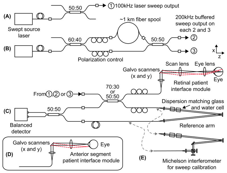
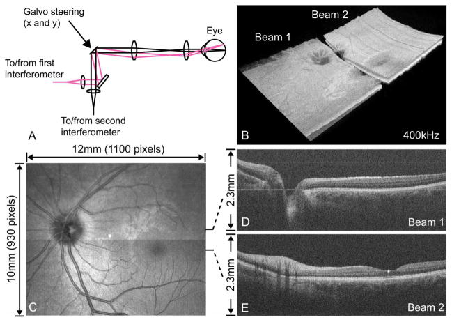
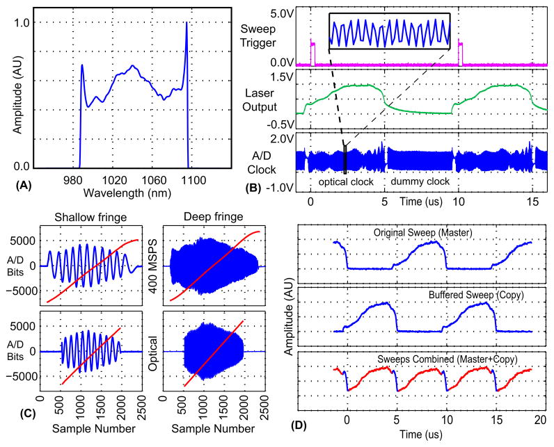
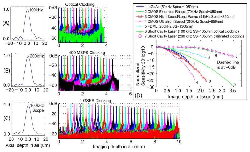
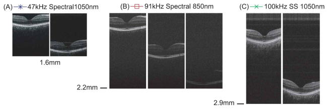
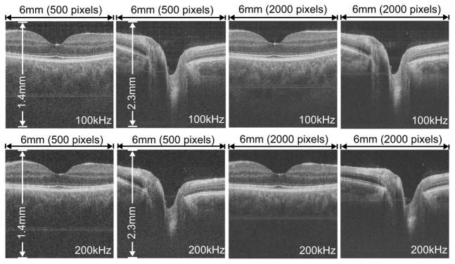
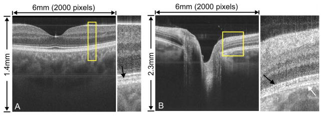
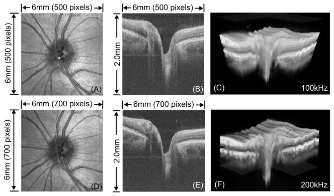
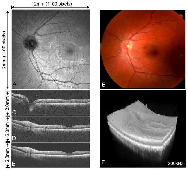
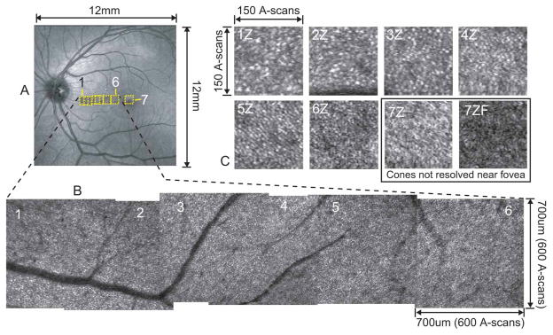
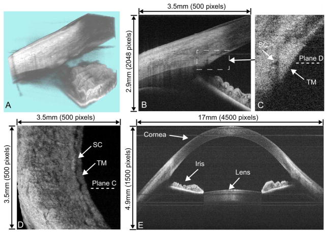
We demonstrate ultrahigh speed swept source/Fourier domain ophthalmic OCT imaging using a short cavity swept laser at 100,000 - 400,000 axial scan rates. Several design configurations illustrate tradeoffs in imaging speed, sensitivity, axial resolution, and imaging depth. Variable rate A/D optical clocking is used to acquire linear-in-k OCT fringe data at 100 kHz axial scan rate with 5.3 um axial resolution in tissue. Fixed rate sampling at 1 GSPS achieves a 7.5mm imaging range in tissue with 6.0 um axial resolution at 100 kHz axial scan rate. A 200 kHz axial scan rate with 5.3 um axial resolution over 4mm imaging range is achieved by buffering the laser sweep. Dual spot OCT using two parallel interferometers achieves 400 kHz axial scan rate, almost 2X faster than previous 1050 nm ophthalmic results and 20X faster than current commercial instruments. Superior sensitivity roll-off performance is shown. Imaging is demonstrated in the human retina and anterior segment. Wide field 12x12 mm data sets include the macula and optic nerve head. Small area, high density imaging shows individual cone photoreceptors. The 7.5 mm imaging range configuration can show the cornea, iris, and anterior lens in a single image. These improvements in imaging speed and depth range provide important advantages for ophthalmic imaging. The ability to rapidly acquire 3D-OCT data over a wide field of view promises to simplify examination protocols. The ability to image fine structures can provide detailed information on focal pathologies. The large imaging range and improved image penetration at 1050 m wavelengths promises to improve performance for instrumentation which images both the retina and anterior eye. These advantages suggest that swept source OCT at 1050 nm wavelengths will play an important role in future ophthalmic instrumentation.
我们展示了使用短腔扫频激光器以100,000 - 400,000轴向扫描速率进行的超高速扫频源/傅里叶域眼科光学相干断层扫描(OCT)成像。几种设计配置说明了成像速度、灵敏度、轴向分辨率和成像深度之间的权衡。可变速率A/D光学时钟用于在组织中以100 kHz轴向扫描速率采集线性k空间OCT条纹数据,轴向分辨率为5.3微米。在1 GSPS的固定速率采样下,在组织中以100 kHz轴向扫描速率实现了7.5毫米的成像范围,轴向分辨率为6.0微米。通过缓冲激光扫描,在4毫米成像范围内实现了200 kHz轴向扫描速率和5.3微米轴向分辨率。使用两个平行干涉仪的双光斑OCT实现了400 kHz轴向扫描速率,几乎比之前1050纳米眼科成像结果快2倍,比当前商业仪器快20倍。展示了卓越的灵敏度滚降性能。在人视网膜和眼前节进行了成像演示。12×12毫米的宽视野数据集包括黄斑和视神经乳头。小区域、高密度成像显示了单个视锥光感受器。7.5毫米成像范围配置可以在单个图像中显示角膜、虹膜和晶状体前部。这些成像速度和深度范围的改进为眼科成像提供了重要优势。在宽视野上快速采集三维OCT数据的能力有望简化检查方案。对精细结构成像的能力可以提供关于局灶性病变的详细信息。在1050纳米波长下大成像范围和改进的图像穿透能力有望提高对视网膜和眼前部进行成像的仪器的性能。这些优势表明,1050纳米波长的扫频源OCT将在未来眼科仪器中发挥重要作用。