Experimental and Clinical Research Center, Charité - University Medicine Berlin, Charitéplatz 1, 10117 Berlin, Germany.
J Neuroinflammation. 2010 Oct 18;7:70. doi: 10.1186/1742-2094-7-70.
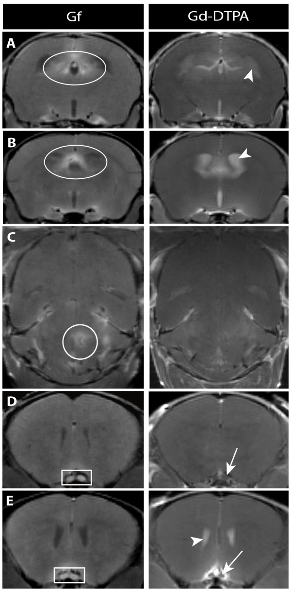
Circumventricular organs (CVO) are cerebral areas with incomplete endothelial blood-brain barrier (BBB) and therefore regarded as "gates to the brain". During inflammation, they may exert an active role in determining immune cell recruitment into the brain.
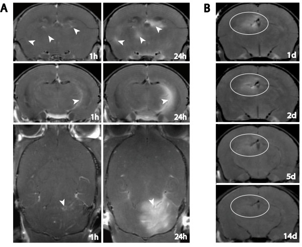
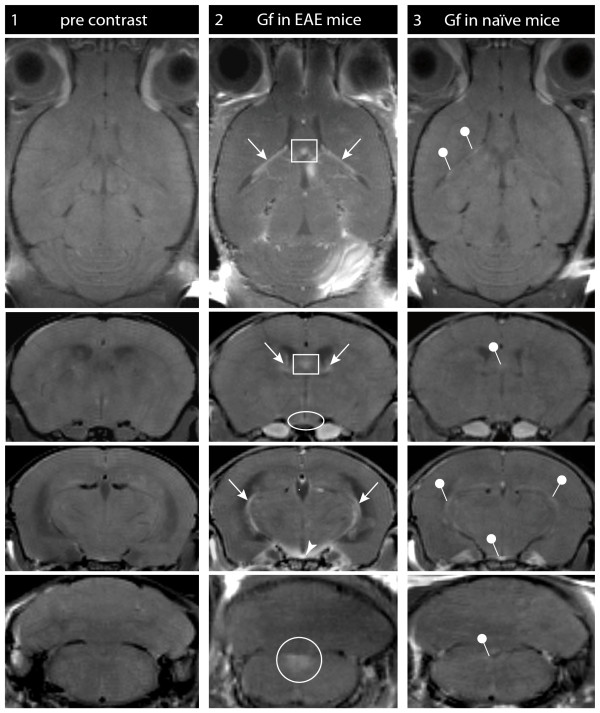
In a longitudinal study we investigated in vivo alterations of CVO during neuroinflammation, applying Gadofluorine M- (Gf) enhanced magnetic resonance imaging (MRI) in experimental autoimmune encephalomyelitis, an animal model of multiple sclerosis. SJL/J mice were monitored by Gadopentate dimeglumine- (Gd-DTPA) and Gf-enhanced MRI after adoptive transfer of proteolipid-protein-specific T cells. Mean Gf intensity ratios were calculated individually for different CVO and correlated to the clinical disease course. Subsequently, the tissue distribution of fluorescence-labeled Gf as well as the extent of cellular inflammation was assessed in corresponding histological slices.
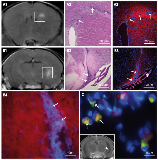
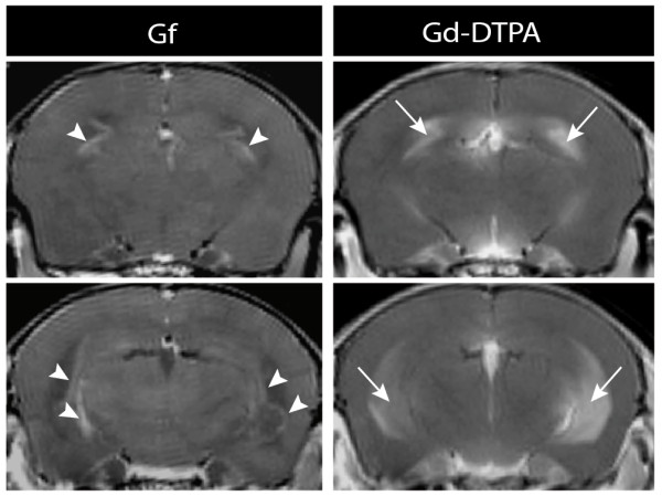
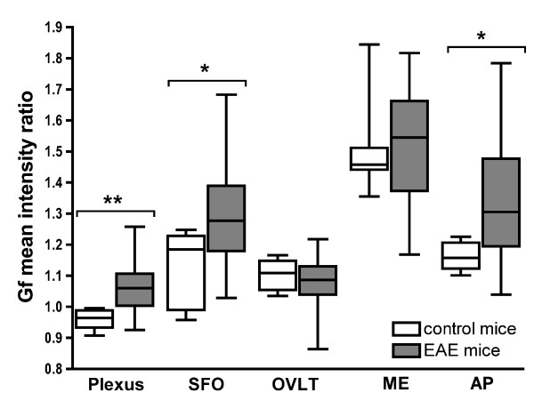
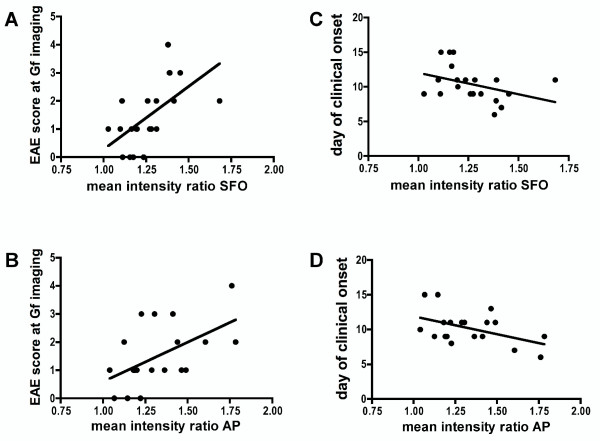
We could show that the Gf signal intensity of the choroid plexus, the subfornicular organ and the area postrema increased significantly during experimental autoimmune encephalomyelitis, correlating with (1) disease severity and (2) the delay of disease onset after immunization. For the choroid plexus, the extent of Gf enhancement served as a diagnostic criterion to distinguish between diseased and healthy control mice with a sensitivity of 89% and a specificity of 80%. Furthermore, Gf improved the detection of lesions, being particularly sensitive to optic neuritis. In correlated histological slices, Gf initially accumulated in the extracellular matrix surrounding inflammatory foci and was subsequently incorporated by macrophages/microglia.
Gf-enhanced MRI provides a novel highly sensitive technique to study cerebral BBB alterations. We demonstrate for the first time in vivo the involvement of CVO during the development of neuroinflammation.
室周器官(CVO)是脑内具有不完全血脑屏障(BBB)的区域,因此被认为是“大脑的门户”。在炎症期间,它们可能在决定免疫细胞向大脑募集方面发挥积极作用。
在一项纵向研究中,我们通过实验性自身免疫性脑脊髓炎(多发性硬化症的动物模型)中的钆氟化物 M(Gf)增强磁共振成像(MRI),研究了神经炎症期间 CVO 的体内变化。在过继转移蛋白脂蛋白蛋白特异性 T 细胞后,用钆喷替酸二甲葡胺(Gd-DTPA)和 Gf 增强 MRI 监测 SJL/J 小鼠。分别计算不同 CVO 的平均 Gf 强度比,并将其与临床疾病过程相关联。随后,在相应的组织切片中评估荧光标记的 Gf 的组织分布和细胞炎症的程度。
我们可以证明,在实验性自身免疫性脑脊髓炎期间,脉络丛、穹窿下器官和后极区的 Gf 信号强度显著增加,这与(1)疾病严重程度和(2)免疫后疾病发作的延迟相关。对于脉络丛,Gf 增强的程度可作为区分患病和健康对照小鼠的诊断标准,其灵敏度为 89%,特异性为 80%。此外,Gf 提高了病变的检测能力,对视神经炎尤为敏感。在相关的组织切片中,Gf 最初积聚在围绕炎症灶的细胞外基质中,随后被巨噬细胞/小胶质细胞摄取。
Gf 增强 MRI 提供了一种研究脑 BBB 变化的新型高灵敏度技术。我们首次在体内证明了 CVO 在神经炎症发展过程中的参与。