Wang Shuangqing, Millward Jason M, Hanke-Vela Laura, Malla Bimala, Pilch Kjara, Gil-Infante Ana, Waiczies Sonia, Mueller Susanne, Boehm-Sturm Philipp, Guo Jing, Sack Ingolf, Infante-Duarte Carmen
Charité - Universitätsmedizin Berlin, Corporate Member of Freie Universität Berlin, Humboldt-Universität zu Berlin, and Berlin Institute of Health, Institute for Medical Immunology, Berlin, Germany.
Department of Neurology, Shenzhen University General Hospital, Shenzhen University Clinical Medical Academy, Shenzhen, China.
Front Neurol. 2020 Jan 13;10:1382. doi: 10.3389/fneur.2019.01382. eCollection 2019.
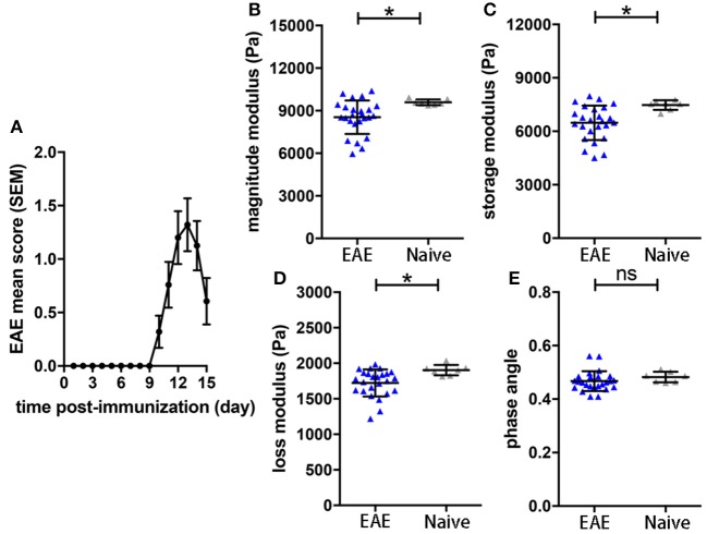
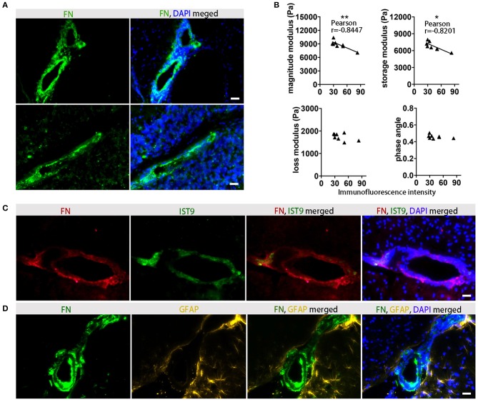
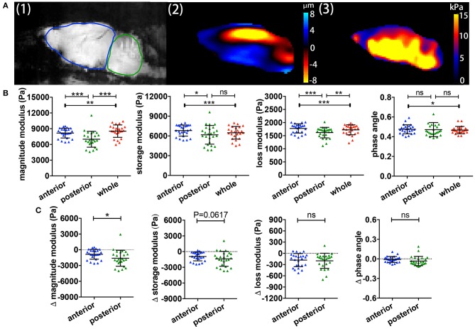
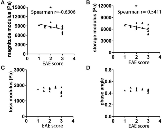
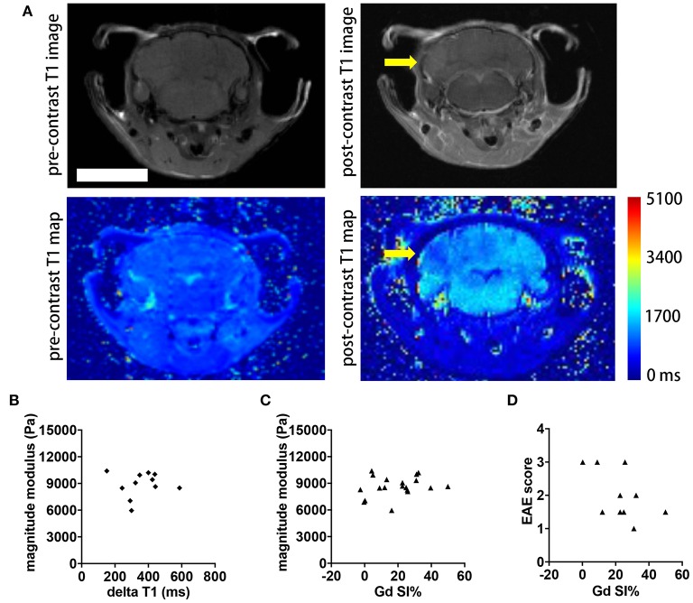
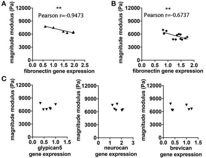
Magnetic resonance imaging (MRI) with gadolinium based contrast agents (GBCA) is routinely used in the clinic to visualize lesions in multiple sclerosis (MS). Although GBCA reveal endothelial permeability, they fail to expose other aspects of lesion formation such as the magnitude of inflammation or tissue changes occurring at sites of blood-brain barrier (BBB) disruption. Moreover, evidence pointing to potential side effects of GBCA has been increasing. Thus, there is an urgent need to develop GBCA-independent imaging tools to monitor pathology in MS. Using MR-elastography (MRE), we previously demonstrated in both MS and the animal model experimental autoimmune encephalomyelitis (EAE) that inflammation was associated with a reduction of brain stiffness. Now, using the relapsing-remitting EAE model, we show that the cerebellum-a region with predominant inflammation in this model-is especially prone to loss of stiffness. We also demonstrate that, contrary to GBCA-MRI, reduction of brain stiffness correlates with clinical disability and is associated with enhanced expression of the extracellular matrix protein fibronectin (FN). Further, we show that FN is largely expressed by activated astrocytes at acute lesions, and reflects the magnitude of tissue remodeling at sites of BBB breakdown. Therefore, MRE could emerge as a safe tool suitable to monitor disease activity in MS.
使用基于钆的造影剂(GBCA)的磁共振成像(MRI)在临床上常用于可视化多发性硬化症(MS)中的病变。尽管GBCA可显示内皮通透性,但它们无法揭示病变形成的其他方面,如炎症程度或血脑屏障(BBB)破坏部位发生的组织变化。此外,指向GBCA潜在副作用的证据一直在增加。因此,迫切需要开发不依赖GBCA的成像工具来监测MS中的病理情况。我们之前使用磁共振弹性成像(MRE)在MS和动物模型实验性自身免疫性脑脊髓炎(EAE)中均证明,炎症与脑硬度降低有关。现在,使用复发缓解型EAE模型,我们表明小脑——该模型中炎症占主导的区域——特别容易出现硬度丧失。我们还证明,与GBCA-MRI相反,脑硬度降低与临床残疾相关,并且与细胞外基质蛋白纤连蛋白(FN)的表达增强有关。此外,我们表明FN在急性病变处主要由活化的星形胶质细胞表达,并反映BBB破坏部位的组织重塑程度。因此,MRE可能成为一种适合监测MS疾病活动的安全工具。