Department of Electrical and Computer Engineering, Missouri University of Science and Technology, Rolla, MO, USA.
Comput Med Imaging Graph. 2011 Mar;35(2):116-20. doi: 10.1016/j.compmedimag.2010.09.006. Epub 2010 Oct 20.
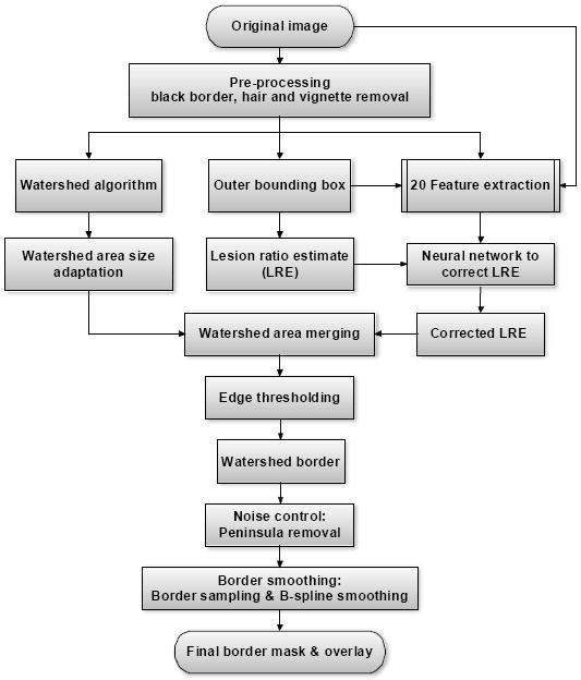
In previous research, a watershed-based algorithm was shown to be useful for automatic lesion segmentation in dermoscopy images, and was tested on a set of 100 benign and malignant melanoma images with the average of three sets of dermatologist-drawn borders used as the ground truth, resulting in an overall error of 15.98%. In this study, to reduce the border detection errors, a neural network classifier was utilized to improve the first-pass watershed segmentation; a novel "edge object value (EOV) threshold" method was used to remove large light blobs near the lesion boundary; and a noise removal procedure was applied to reduce the peninsula-shaped false-positive areas. As a result, an overall error of 11.09% was achieved.
在之前的研究中,基于分水岭的算法被证明对皮肤镜图像的自动病变分割很有用,并在一组 100 张良性和恶性黑色素瘤图像上进行了测试,平均使用三组皮肤科医生绘制的边界作为金标准,整体错误率为 15.98%。在这项研究中,为了减少边界检测误差,利用神经网络分类器改进了初次分水岭分割;采用了一种新的“边缘对象值(EOV)阈值”方法来去除病变边界附近的大光斑;并应用了噪声去除过程来减少半岛状的假阳性区域。结果,整体错误率达到了 11.09%。