International Centre for Eye Health, Department of Infectious and Tropical Diseases, London School of Hygiene and Tropical Medicine, London, United Kingdom.
Ophthalmology. 2011 Apr;118(4):747-54. doi: 10.1016/j.ophtha.2010.08.029. Epub 2010 Nov 5.
To describe the in vivo confocal microscopy (IVCM) appearances of the tarsal conjunctiva in trachoma compared with the appearance of healthy conjunctiva and to develop grading systems for IVCM examination of the tarsal conjunctiva for use in future studies on trachoma and other conjunctival diseases.
Prospective observational study.
In vivo confocal microscopy examination was performed on 302 clinically normal adults, 16 clinically normal children, 750 adults with trachomatous conjunctival scarring, and 25 children with active trachoma.
Clinical evaluation was performed with ×2.5 loupes, and IVCM examination of the upper tarsal conjunctiva was carried out with a Heidelberg Retina Tomograph 3 with the Rostock Cornea Module (Heidelberg Engineering GmbH, Dossenheim, Germany).
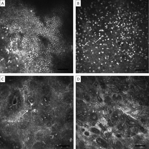
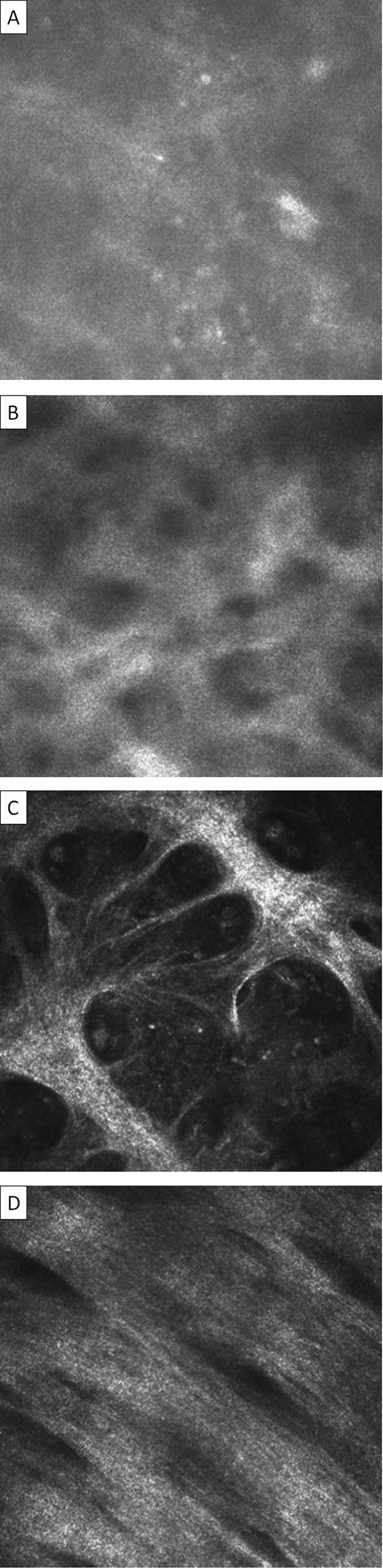
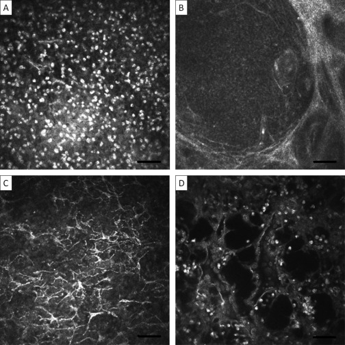
In vivo confocal microscopy images were analyzed for cellular and tissue changes associated with trachomatous inflammation and scarring compared with healthy subjects.
Trachomatous subjects with follicular and papillary inflammation had an increased inflammatory cellular infiltrate, including dendritiform cells, discrete follicular and papillary structures, and cystic lacunae suggestive of tissue edema. Trachomatous conjunctival scarring was seen with IVCM as organization of the subepithelial connective tissue into bands/sheets. Grading systems for inflammatory changes and scarring were developed, with the system for scarring showing good interobserver agreement with an intraclass coefficient of 0.88.
In vivo confocal microscopy provides a powerful tool for examining the ocular surface. Numerous cellular and tissue changes were observed in subjects with trachoma, the first time IVCM has been applied to this disease. These changes both complement and add to previous histologic analyses. In vivo confocal microscopy promises to provide new insights into the pathogenesis of trachoma and other conjunctival diseases.
描述沙眼患者的睑结膜活体共聚焦显微镜(IVCM)表现,并与健康结膜的表现进行比较,同时制定用于未来沙眼及其他结膜疾病研究的睑结膜 IVCM 检查分级系统。
前瞻性观察性研究。
对 302 名临床正常成年人、16 名临床正常儿童、750 名患有沙眼性结膜瘢痕的成年人和 25 名患有活动性沙眼的儿童进行了 IVCM 检查。
使用 ×2.5 倍放大镜进行临床评估,使用海德堡视网膜断层扫描仪 3 号与罗斯托克角膜模块(海德堡工程公司,德国多森海姆)进行上睑结膜 IVCM 检查。
与健康受试者相比,分析与沙眼炎症和瘢痕相关的细胞和组织变化的活体共聚焦显微镜图像。
滤泡性和乳头状炎症的沙眼患者存在炎症细胞浸润增加,包括树突状细胞、离散的滤泡和乳头状结构以及提示组织水肿的囊性腔隙。IVCM 可观察到沙眼性结膜瘢痕,表现为基质下结缔组织呈带/片状排列。建立了炎症改变和瘢痕的分级系统,瘢痕分级系统的观察者间一致性良好,组内相关系数为 0.88。
活体共聚焦显微镜为检查眼表面提供了强有力的工具。在沙眼患者中观察到许多细胞和组织变化,这是首次将 IVCM 应用于该疾病。这些变化既补充又补充了之前的组织学分析。活体共聚焦显微镜有望为沙眼和其他结膜疾病的发病机制提供新的见解。