• 文献检索
  • 文档翻译
  • 深度研究
  • 学术资讯
  • Suppr Zotero 插件Zotero 插件
  • 邀请有礼
  • 套餐&价格
  • 历史记录
应用&插件
Suppr Zotero 插件Zotero 插件浏览器插件Mac 客户端Windows 客户端微信小程序
定价
高级版会员购买积分包购买API积分包
服务
文献检索文档翻译深度研究API 文档MCP 服务
关于我们
关于 Suppr公司介绍联系我们用户协议隐私条款
关注我们

Suppr 超能文献

核心技术专利:CN118964589B侵权必究
粤ICP备2023148730 号-1Suppr @ 2026

文献检索

告别复杂PubMed语法,用中文像聊天一样搜索,搜遍4000万医学文献。AI智能推荐,让科研检索更轻松。

立即免费搜索

文件翻译

保留排版,准确专业,支持PDF/Word/PPT等文件格式,支持 12+语言互译。

免费翻译文档

深度研究

AI帮你快速写综述,25分钟生成高质量综述,智能提取关键信息,辅助科研写作。

立即免费体验

沙眼瘢痕和正常组织的结膜共焦显微镜和组织病理学的体内比较:系统比较。

In vivo confocal microscopy and histopathology of the conjunctiva in trachomatous scarring and normal tissue: a systematic comparison.

机构信息

Faculty of Infectious and Tropical Diseases, International Centre for Eye Health, London School of Hygiene and Tropical Medicine, , London, UK.

出版信息

Br J Ophthalmol. 2013 Oct;97(10):1333-7. doi: 10.1136/bjophthalmol-2013-303126. Epub 2013 Aug 6.

DOI:10.1136/bjophthalmol-2013-303126
PMID:23922266
原文链接:https://pmc.ncbi.nlm.nih.gov/articles/PMC3786664/
Abstract

AIM

To compare in vivo confocal microscopy (IVCM) with the histopathological examination of tissue and cellular changes in normal and diseased conjunctiva.

METHODS

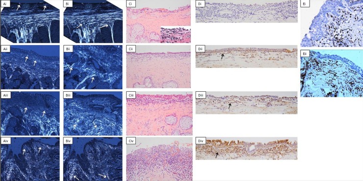
Participants underwent clinical examination and IVCM of the tarsal conjunctiva. A biopsy of the upper tarsal conjunctiva was collected and stained with tinctorial stains and by immunohistochemical staining for CD45 and CD83. Connective tissue scarring, inflammatory cell density and the presence of dendritiform cells were quantitatively assessed in a masked manner by both IVCM and histological assessments for comparative analysis.

RESULTS

Thirty-four participants with severe trachomatous conjunctival scarring and 33 participants with healthy conjunctiva were recruited. The IVCM connective tissue scarring score was strongly associated with the histological grading of scarring (p<0.001). There was limited evidence of an association between the IVCM inflammatory cell infiltrate and the histological inflammatory cell grade (p=0.05). We did not find any evidence to support the hypothesis that dendritiform cells seen with IVCM are mature, conventional dendritic cells.

CONCLUSIONS

The results show that IVCM can be used to robustly quantitate connective tissue scarring and also has a role in measuring the inflammatory cell infiltrate. The discordance between IVCM dendritiform cells and immunohistochemical dendritic cells may be a result of study limitations or may be because these dendritiform structures represent another cell type, such as fibroblasts, rather than dendritic cells.

摘要

目的

比较活体共聚焦显微镜(IVCM)与组织和细胞在正常和病变结膜中的组织病理学检查。

方法

参与者接受了睑结膜的临床检查和 IVCM。收集上睑结膜活检标本,并用染色剂和 CD45 和 CD83 的免疫组织化学染色进行染色。通过 IVCM 和组织学评估以盲法对结缔组织瘢痕、炎性细胞密度和树突状细胞的存在进行定量评估,并进行比较分析。

结果

招募了 34 名严重的沙眼性结膜瘢痕患者和 33 名健康结膜患者。IVCM 结缔组织瘢痕评分与组织学瘢痕分级密切相关(p<0.001)。IVCM 炎性细胞浸润与组织学炎性细胞分级之间的关联证据有限(p=0.05)。我们没有发现任何证据支持 IVCM 中观察到的树突状细胞是成熟的、常规树突状细胞的假设。

结论

结果表明,IVCM 可用于稳健地定量评估结缔组织瘢痕,并且在测量炎性细胞浸润方面也有作用。IVCM 中的树突状细胞与免疫组织化学树突状细胞之间的不相符可能是由于研究限制所致,或者可能是因为这些树突状结构代表另一种细胞类型,例如成纤维细胞,而不是树突状细胞。

https://cdn.ncbi.nlm.nih.gov/pmc/blobs/a68f/3786664/4f605aa17628/bjophthalmol-2013-303126f01.jpg

相似文献

1
In vivo confocal microscopy and histopathology of the conjunctiva in trachomatous scarring and normal tissue: a systematic comparison.沙眼瘢痕和正常组织的结膜共焦显微镜和组织病理学的体内比较:系统比较。
Br J Ophthalmol. 2013 Oct;97(10):1333-7. doi: 10.1136/bjophthalmol-2013-303126. Epub 2013 Aug 6.
2
In vivo confocal microscopy in scarring trachoma.在瘢痕性沙眼的活体共聚焦显微镜检查。
Ophthalmology. 2011 Nov;118(11):2138-46. doi: 10.1016/j.ophtha.2011.04.014. Epub 2011 Sep 13.
3
In vivo confocal microscopy of trachoma in relation to normal tarsal conjunctiva.沙眼的活体共聚焦显微镜检查与正常睑结膜的关系。
Ophthalmology. 2011 Apr;118(4):747-54. doi: 10.1016/j.ophtha.2010.08.029. Epub 2010 Nov 5.
4
Immunohistochemical Analysis of Scarring Trachoma Indicates Infiltration by Natural Killer and Undefined CD45 Negative Cells.瘢痕性沙眼的免疫组织化学分析表明存在自然杀伤细胞和未明确的CD45阴性细胞浸润。
PLoS Negl Trop Dis. 2016 May 24;10(5):e0004734. doi: 10.1371/journal.pntd.0004734. eCollection 2016 May.
5
In vivo confocal microscopy and trachomatous conjunctival scarring: Predictors for clinical progression.体内共聚焦显微镜和沙眼性结膜瘢痕:临床进展的预测因素。
Clin Exp Ophthalmol. 2020 Dec;48(9):1152-1159. doi: 10.1111/ceo.13843. Epub 2020 Sep 16.
6
Innate immune responses and modified extracellular matrix regulation characterize bacterial infection and cellular/connective tissue changes in scarring trachoma.先天免疫反应和细胞外基质调节的改变是致瘢痕性沙眼的细菌感染和细胞/结缔组织改变的特征。
Infect Immun. 2012 Jan;80(1):121-30. doi: 10.1128/IAI.05965-11. Epub 2011 Oct 28.
7
Pathogenesis of progressive scarring trachoma in Ethiopia and Tanzania and its implications for disease control: two cohort studies.埃塞俄比亚和坦桑尼亚进行性瘢痕性沙眼的发病机制及其对疾病控制的意义:两项队列研究
PLoS Negl Trop Dis. 2015 May 13;9(5):e0003763. doi: 10.1371/journal.pntd.0003763. eCollection 2015 May.
8
Diagnosing ocular surface squamous neoplasia in East Africa: case-control study of clinical and in vivo confocal microscopy assessment.在东非诊断眼表鳞状上皮肿瘤:临床与活体共聚焦显微镜评估的病例对照研究。
Ophthalmology. 2014 Feb;121(2):484-91. doi: 10.1016/j.ophtha.2013.09.027. Epub 2013 Dec 8.
9
Conjunctival scarring in trachoma is associated with the HLA-C ligand of KIR and is exacerbated by heterozygosity at KIR2DL2/KIR2DL3.沙眼的结膜瘢痕与 KIR 的 HLA-C 配体有关,并且 KIR2DL2/KIR2DL3 的杂合性会使其恶化。
PLoS Negl Trop Dis. 2014 Mar 20;8(3):e2744. doi: 10.1371/journal.pntd.0002744. eCollection 2014 Mar.
10
In Vivo Confocal Microscopy in Dry Eye Disease Associated With Chronic Graft-Versus-Host Disease.与慢性移植物抗宿主病相关的干眼病的体内共聚焦显微镜检查
Invest Ophthalmol Vis Sci. 2016 Sep 1;57(11):4686-91. doi: 10.1167/iovs.16-20013.

引用本文的文献

1
A review of the application of in-vivo confocal microscopy on conjunctival diseases.共聚焦显微镜在结膜疾病中的应用综述
Eye Vis (Lond). 2024 Nov 1;11(1):43. doi: 10.1186/s40662-024-00409-x.
2
Insights into Pathogenesis of Trachoma.沙眼发病机制的见解
Microorganisms. 2024 Jul 28;12(8):1544. doi: 10.3390/microorganisms12081544.
3
Slit lamp polarized dermoscopy: a cost-effective tool to assess eyelid lesions.裂隙灯偏光皮肤镜检查:评估眼睑病变的一种具有成本效益的工具。

本文引用的文献

1
Innate immune responses and modified extracellular matrix regulation characterize bacterial infection and cellular/connective tissue changes in scarring trachoma.先天免疫反应和细胞外基质调节的改变是致瘢痕性沙眼的细菌感染和细胞/结缔组织改变的特征。
Infect Immun. 2012 Jan;80(1):121-30. doi: 10.1128/IAI.05965-11. Epub 2011 Oct 28.
2
In vivo confocal microscopy in scarring trachoma.在瘢痕性沙眼的活体共聚焦显微镜检查。
Ophthalmology. 2011 Nov;118(11):2138-46. doi: 10.1016/j.ophtha.2011.04.014. Epub 2011 Sep 13.
3
Diagnostic value of confocal laser endomicroscopy for gastric superficial cancerous lesions.
Int Ophthalmol. 2023 Apr;43(4):1103-1110. doi: 10.1007/s10792-022-02505-0. Epub 2022 Sep 9.
4
Trachoma.沙眼。
Nat Rev Dis Primers. 2022 May 26;8(1):32. doi: 10.1038/s41572-022-00359-5.
5
Primary conjunctival tuberculosis in two middle-aged women.两名中年女性的原发性结膜结核
Int J Ophthalmol. 2020 Jan 18;13(1):180-183. doi: 10.18240/ijo.2020.01.25. eCollection 2020.
6
Immunofibrogenic Gene Expression Patterns in Tanzanian Children with Ocular Infection, Active Trachoma and Scarring: Baseline Results of a 4-Year Longitudinal Study.坦桑尼亚儿童眼部感染、活动性沙眼和瘢痕形成的免疫纤维生成基因表达模式:一项为期 4 年的纵向研究的基线结果。
Front Cell Infect Microbiol. 2017 Sep 15;7:406. doi: 10.3389/fcimb.2017.00406. eCollection 2017.
7
Fibroblasts profiling in scarring trachoma identifies IL-6 as a functional component of a fibroblast-macrophage pro-fibrotic and pro-inflammatory feedback loop.瘢痕性沙眼成纤维细胞分析确定白细胞介素-6是成纤维细胞-巨噬细胞促纤维化和促炎反馈回路的功能成分。
Sci Rep. 2016 Jun 20;6:28261. doi: 10.1038/srep28261.
8
Increased Epithelial Expression of CTGF and S100A7 with Elevated Subepithelial Expression of IL-1β in Trachomatous Trichiasis.沙眼性倒睫中,CTGF和S100A7的上皮表达增加,同时IL-1β的上皮下表达升高。
PLoS Negl Trop Dis. 2016 Jun 1;10(6):e0004752. doi: 10.1371/journal.pntd.0004752. eCollection 2016 Jun.
9
Immunohistochemical Analysis of Scarring Trachoma Indicates Infiltration by Natural Killer and Undefined CD45 Negative Cells.瘢痕性沙眼的免疫组织化学分析表明存在自然杀伤细胞和未明确的CD45阴性细胞浸润。
PLoS Negl Trop Dis. 2016 May 24;10(5):e0004734. doi: 10.1371/journal.pntd.0004734. eCollection 2016 May.
10
Trachoma and Ocular Chlamydial Infection in the Era of Genomics.基因组学时代的沙眼和眼部衣原体感染
Mediators Inflamm. 2015;2015:791847. doi: 10.1155/2015/791847. Epub 2015 Sep 3.
共聚焦激光内镜检查对胃表浅癌性病变的诊断价值。
Gut. 2011 Mar;60(3):299-306. doi: 10.1136/gut.2010.223586. Epub 2010 Dec 30.
4
Bacterial infection in scarring trachoma.瘢痕性沙眼的细菌感染。
Invest Ophthalmol Vis Sci. 2011 Apr 6;52(5):2181-6. doi: 10.1167/iovs.10-5829. Print 2011 Apr.
5
In vivo confocal microscopy of trachoma in relation to normal tarsal conjunctiva.沙眼的活体共聚焦显微镜检查与正常睑结膜的关系。
Ophthalmology. 2011 Apr;118(4):747-54. doi: 10.1016/j.ophtha.2010.08.029. Epub 2010 Nov 5.
6
In vivo confocal microscopic evaluation of morphologic changes and dendritic cell distribution in pterygium.翼状胬肉中形态变化和树突状细胞分布的体内共聚焦显微镜评估。
Am J Ophthalmol. 2010 Nov;150(5):650-655.e1. doi: 10.1016/j.ajo.2010.05.025. Epub 2010 Aug 5.
7
In vivo laser scanning confocal microscopy of vernal keratoconjunctivitis.春季角结膜炎的活体激光共聚焦显微镜检查。
Clin Exp Ophthalmol. 2011 Jan;39(1):53-60. doi: 10.1111/j.1442-9071.2010.02379.x.
8
[Reflectance confocal microscopy in the diagnosis of cutaneous melanoma].[反射式共聚焦显微镜在皮肤黑色素瘤诊断中的应用]
An Bras Dermatol. 2009 Nov-Dec;84(6):636-42. doi: 10.1590/s0365-05962009000600009.
9
Clinical in vivo confocal microscopy of the human cornea in health and disease.健康和疾病人群角膜的临床活体共聚焦显微镜检查。
Prog Retin Eye Res. 2010 Jan;29(1):30-58. doi: 10.1016/j.preteyeres.2009.11.001. Epub 2009 Nov 26.
10
Origin, homeostasis and function of Langerhans cells and other langerin-expressing dendritic cells.朗格汉斯细胞及其他表达朗格琳的树突状细胞的起源、稳态与功能。
Nat Rev Immunol. 2008 Dec;8(12):935-47. doi: 10.1038/nri2455.