Division of Pulmonary and Critical Care Medicine, Department of Medicine, Samsung Medical Center, Sungkyunkwan University School of Medicine, Seoul, Korea.
Korean J Radiol. 2010 Nov-Dec;11(6):612-7. doi: 10.3348/kjr.2010.11.6.612. Epub 2010 Oct 29.
To describe the radiographic findings of primary pulmonary tuberculosis (TB) in previously healthy adolescent patients.
The Institutional Review Board approved this retrospective study, with a waiver of informed consent from the patients. TB outbreaks occurred in 15 senior high schools and chest radiographs from 58 students with identical strains of TB were analyzed by restriction fragment length polymorphism analysis by two independent observers. Lesions of nodule(s), consolidation, or cavitation in the upper lung zones were classified as typical TB. Mediastinal lymph node enlargement; lesions of nodule(s), consolidation, or cavitation in lower lung zones; or pleural effusion were classified as atypical TB. Inter-observer agreement for the presence of each radiographic finding was examined by kappa statistics.
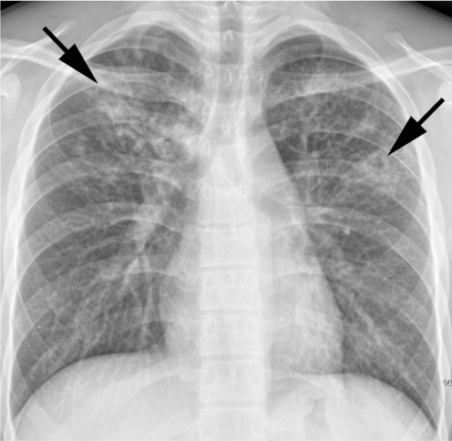
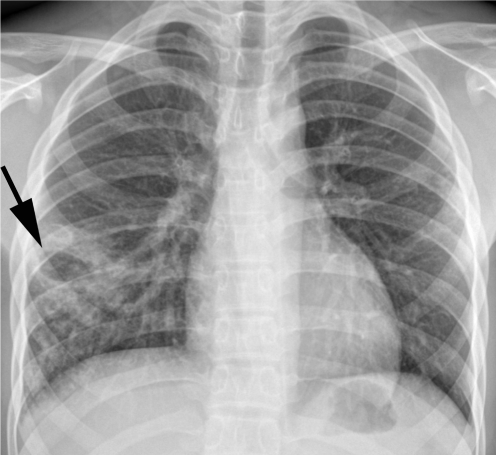
Of 58 patients, three (5%) had normal chest radiographs. Cavitary lesions were present in 25 (45%) of 55 students. Lesions with upper lung zone predominance were observed in 27 (49%) patients, whereas lower lung zone predominance was noted in 18 (33%) patients. The remaining 10 (18%) patients had lesions in both upper and lower lung zones. Pleural effusion was not observed in any patient, nor was the mediastinal lymph node enlargement. Hilar lymph node enlargement was seen in only one (2%) patient. Overall, 37 (67%) students had the typical form of TB, whereas 18 (33%) had TB lesions of the atypical form.
The most common radiographic findings in primary pulmonary TB by recent infection in previously healthy adolescents are upper lung lesions, which were thought to be radiographic findings of reactivation pulmonary TB by remote infection.
描述既往健康的青少年患者原发性肺结核(TB)的放射学表现。
本回顾性研究经机构审查委员会批准,患者免除了知情同意。15 所高级中学发生了结核病爆发,由两位独立观察者通过限制性片段长度多态性分析对具有相同结核菌株的 58 名学生的胸部 X 线片进行了分析。上肺区的结节、实变或空洞病变被归类为典型 TB。纵隔淋巴结肿大;下肺区的结节、实变或空洞病变;或胸腔积液被归类为非典型 TB。通过 Kappa 统计检验评估每个放射学表现的观察者间一致性。
58 例患者中,3 例(5%)胸部 X 线片正常。55 例学生中有 25 例(45%)有空洞病变。27 例(49%)患者表现为上肺区病变为主,18 例(33%)患者表现为下肺区病变为主。其余 10 例(18%)患者有上、下肺区病变。没有患者出现胸腔积液,也没有纵隔淋巴结肿大。仅 1 例(2%)患者有肺门淋巴结肿大。总体而言,37 例(67%)学生有典型的 TB 表现,18 例(33%)学生有非典型的 TB 病变。
既往健康的青少年近期感染原发性肺结核的最常见放射学表现为上肺病变,这些病变被认为是既往感染引起的再激活性肺结核的放射学表现。