• 文献检索
  • 文档翻译
  • 深度研究
  • 学术资讯
  • Suppr Zotero 插件Zotero 插件
  • 邀请有礼
  • 套餐&价格
  • 历史记录
应用&插件
Suppr Zotero 插件Zotero 插件浏览器插件Mac 客户端Windows 客户端微信小程序
定价
高级版会员购买积分包购买API积分包
服务
文献检索文档翻译深度研究API 文档MCP 服务
关于我们
关于 Suppr公司介绍联系我们用户协议隐私条款
关注我们

Suppr 超能文献

核心技术专利:CN118964589B侵权必究
粤ICP备2023148730 号-1Suppr @ 2026

文献检索

告别复杂PubMed语法,用中文像聊天一样搜索,搜遍4000万医学文献。AI智能推荐,让科研检索更轻松。

立即免费搜索

文件翻译

保留排版,准确专业,支持PDF/Word/PPT等文件格式,支持 12+语言互译。

免费翻译文档

深度研究

AI帮你快速写综述,25分钟生成高质量综述,智能提取关键信息,辅助科研写作。

立即免费体验

小分子亲水型内质网相关蛋白(SHERP)在细胞膜表面的分子识别的结构基础。

Structural basis of molecular recognition of the Leishmania small hydrophilic endoplasmic reticulum-associated protein (SHERP) at membrane surfaces.

机构信息

Division of Cell and Molecular Biology, Centre for Molecular Microbiology and Infection, Imperial College London, Exhibition Road, London SW7 2AZ, United Kingdom.

出版信息

J Biol Chem. 2011 Mar 18;286(11):9246-56. doi: 10.1074/jbc.M110.130427. Epub 2010 Nov 24.

DOI:10.1074/jbc.M110.130427
PMID:21106528
原文链接:https://pmc.ncbi.nlm.nih.gov/articles/PMC3059043/
Abstract

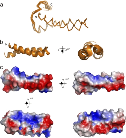
The 57-residue small hydrophilic endoplasmic reticulum-associated protein (SHERP) shows highly specific, stage-regulated expression in the non-replicative vector-transmitted stages of the kinetoplastid parasite, Leishmania major, the causative agent of human cutaneous leishmaniasis. Previous studies have demonstrated that SHERP localizes as a peripheral membrane protein on the cytosolic face of the endoplasmic reticulum and on outer mitochondrial membranes, whereas its high copy number suggests a critical function in vivo. However, the absence of defined domains or identifiable orthologues, together with lack of a clear phenotype in transgenic parasites lacking SHERP, has limited functional understanding of this protein. Here, we use a combination of biophysical and biochemical methods to demonstrate that SHERP can be induced to adopt a globular fold in the presence of anionic lipids or SDS. Cross-linking and binding studies suggest that SHERP has the potential to form a complex with the vacuolar type H(+)-ATPase. Taken together, these results suggest that SHERP may function in modulating cellular processes related to membrane organization and/or acidification during vector transmission of infective Leishmania.

摘要

57 个残基的小亲水性内质网相关蛋白(SHERP)在无复制性载体传播阶段的锥虫寄生虫利什曼原虫中表现出高度特异性、阶段调节表达,该寄生虫是人类皮肤利什曼病的病原体。先前的研究表明,SHERP 作为一种外周膜蛋白定位于内质网的细胞质面和外线粒体膜上,而其高拷贝数表明其在体内具有关键功能。然而,由于缺乏定义的结构域或可识别的同源物,以及缺乏缺乏 SHERP 的转基因寄生虫的明确表型,限制了对这种蛋白质的功能理解。在这里,我们使用生物物理和生化方法的组合来证明,在阴离子脂质或 SDS 的存在下,SHERP 可以被诱导采用球形折叠。交联和结合研究表明,SHERP 有可能与液泡型 H(+)-ATP 酶形成复合物。总之,这些结果表明,SHERP 可能在调节与膜组织和/或酸化相关的细胞过程中发挥作用,这与感染性利什曼原虫的载体传播有关。

https://cdn.ncbi.nlm.nih.gov/pmc/blobs/0d55/3059043/2c8d9b379522/zbc0101148490005.jpg
https://cdn.ncbi.nlm.nih.gov/pmc/blobs/0d55/3059043/d7af712fe7b8/zbc0101148490001.jpg
https://cdn.ncbi.nlm.nih.gov/pmc/blobs/0d55/3059043/426cd76e7283/zbc0101148490002.jpg
https://cdn.ncbi.nlm.nih.gov/pmc/blobs/0d55/3059043/37eedd6eaff8/zbc0101148490003.jpg
https://cdn.ncbi.nlm.nih.gov/pmc/blobs/0d55/3059043/169050047684/zbc0101148490004.jpg
https://cdn.ncbi.nlm.nih.gov/pmc/blobs/0d55/3059043/2c8d9b379522/zbc0101148490005.jpg

相似文献

1
Structural basis of molecular recognition of the Leishmania small hydrophilic endoplasmic reticulum-associated protein (SHERP) at membrane surfaces.小分子亲水型内质网相关蛋白(SHERP)在细胞膜表面的分子识别的结构基础。
J Biol Chem. 2011 Mar 18;286(11):9246-56. doi: 10.1074/jbc.M110.130427. Epub 2010 Nov 24.
2
Characterization of a differentially expressed protein that shows an unusual localization to intracellular membranes in Leishmania major.一种差异表达蛋白的特性研究,该蛋白在硕大利什曼原虫中定位于细胞内膜,表现出异常的定位情况。
Biochem J. 2001 Jun 1;356(Pt 2):335-44. doi: 10.1042/0264-6021:3560335.
3
Detection of Leishmania metacyclogenesis within the sand fly vector employing a real-time PCR for sherp gene expression: A tool for Leishmania surveillance and transmission potential.利用实时PCR检测沙蝇媒介内利什曼原虫的循环前体形成以监测sherp基因表达:一种用于利什曼原虫监测和传播潜力评估的工具。
PLoS Negl Trop Dis. 2025 Mar 17;19(3):e0012915. doi: 10.1371/journal.pntd.0012915. eCollection 2025 Mar.
4
Phenotypic changes associated with deletion and overexpression of a stage-regulated gene family in Leishmania.与利什曼原虫中一个阶段调控基因家族的缺失和过表达相关的表型变化。
Cell Microbiol. 2001 Aug;3(8):511-23. doi: 10.1046/j.1462-5822.2001.00135.x.
5
Leishmania HASP and SHERP Genes Are Required for In Vivo Differentiation, Parasite Transmission and Virulence Attenuation in the Host.利什曼原虫的HASP和SHERP基因是体内分化、寄生虫传播及宿主毒力减弱所必需的。
PLoS Pathog. 2017 Jan 17;13(1):e1006130. doi: 10.1371/journal.ppat.1006130. eCollection 2017 Jan.
6
The stage-regulated HASPB and SHERP proteins are essential for differentiation of the protozoan parasite Leishmania major in its sand fly vector, Phlebotomus papatasi.阶段调控的 HASPB 和 SHERP 蛋白对于原生动物寄生虫利什曼原虫在其沙蝇媒介白蛉中的分化是必不可少的。
Cell Microbiol. 2010 Dec;12(12):1765-79. doi: 10.1111/j.1462-5822.2010.01507.x.
7
Trafficking and release of Leishmania metacyclic HASPB on macrophage invasion.利什曼原虫无鞭毛体在巨噬细胞入侵过程中的转运和释放。
Cell Microbiol. 2012 May;14(5):740-61. doi: 10.1111/j.1462-5822.2012.01756.x. Epub 2012 Feb 24.
8
Disruption of the Putative Ribosome-Binding Motif of a Scaffold Protein Impairs Cytochrome Oxidase Subunit Expression in .支架蛋白的核糖体结合基序缺失会影响. 中的细胞色素氧化酶亚基的表达。
mSphere. 2019 Mar 6;4(2):e00644-18. doi: 10.1128/mSphere.00644-18.
9
Functional genomics in sand fly-derived Leishmania promastigotes.沙蝇源性利什曼原虫的功能基因组学。
PLoS Negl Trop Dis. 2019 May 9;13(5):e0007288. doi: 10.1371/journal.pntd.0007288. eCollection 2019 May.
10
Endoplasmic reticulum stress-induced apoptosis in Leishmania through Ca2+-dependent and caspase-independent mechanism.内质网应激诱导利什曼原虫通过 Ca2+-依赖性和 caspase 非依赖性机制凋亡。
J Biol Chem. 2011 Apr 15;286(15):13638-46. doi: 10.1074/jbc.M110.201889. Epub 2011 Feb 17.

引用本文的文献

1
Detection of Leishmania metacyclogenesis within the sand fly vector employing a real-time PCR for sherp gene expression: A tool for Leishmania surveillance and transmission potential.利用实时PCR检测沙蝇媒介内利什曼原虫的循环前体形成以监测sherp基因表达:一种用于利什曼原虫监测和传播潜力评估的工具。
PLoS Negl Trop Dis. 2025 Mar 17;19(3):e0012915. doi: 10.1371/journal.pntd.0012915. eCollection 2025 Mar.
2
CYP5122A1 encodes an essential sterol C4-methyl oxidase in Leishmania donovani and determines the antileishmanial activity of antifungal azoles.CYP5122A1 编码利什曼原虫中的一种必需固醇 C4-甲基氧化酶,决定了抗真菌唑类药物的抗利什曼原虫活性。
Nat Commun. 2024 Oct 31;15(1):9409. doi: 10.1038/s41467-024-53790-5.
3

本文引用的文献

1
The stage-regulated HASPB and SHERP proteins are essential for differentiation of the protozoan parasite Leishmania major in its sand fly vector, Phlebotomus papatasi.阶段调控的 HASPB 和 SHERP 蛋白对于原生动物寄生虫利什曼原虫在其沙蝇媒介白蛉中的分化是必不可少的。
Cell Microbiol. 2010 Dec;12(12):1765-79. doi: 10.1111/j.1462-5822.2010.01507.x.
2
Structure of intact Thermus thermophilus V-ATPase by cryo-EM reveals organization of the membrane-bound V(O) motor.冷冻电镜解析完整的嗜热栖热菌 V-ATP 酶结构揭示了膜结合 V(O)马达的组装。
Proc Natl Acad Sci U S A. 2010 Jan 26;107(4):1367-72. doi: 10.1073/pnas.0911085107. Epub 2010 Jan 6.
3
The gene expression of Leishmania infantum chagasi inside Lutzomyia longipalpis, the main vector of visceral leishmaniasis in Brazil.
巴西内脏利什曼病主要传播媒介白蛉(Lutzomyia longipalpis)体内查加斯利什曼原虫(Leishmania infantum chagasi)的基因表达。
Mem Inst Oswaldo Cruz. 2021 Mar 8;116:e200571. doi: 10.1590/0074-02760200571. eCollection 2021.
4
Differential dehydration effects on globular proteins and intrinsically disordered proteins during film formation.成膜过程中球状蛋白和内在无序蛋白的脱水差异效应。
Protein Sci. 2017 Apr;26(4):718-726. doi: 10.1002/pro.3118. Epub 2017 Feb 7.
5
Leishmania HASP and SHERP Genes Are Required for In Vivo Differentiation, Parasite Transmission and Virulence Attenuation in the Host.利什曼原虫的HASP和SHERP基因是体内分化、寄生虫传播及宿主毒力减弱所必需的。
PLoS Pathog. 2017 Jan 17;13(1):e1006130. doi: 10.1371/journal.ppat.1006130. eCollection 2017 Jan.
6
Distinct circular dichroism spectroscopic signatures of polyproline II and unordered secondary structures: applications in secondary structure analyses.聚脯氨酸II和无规二级结构独特的圆二色光谱特征:在二级结构分析中的应用。
Protein Sci. 2014 Dec;23(12):1765-72. doi: 10.1002/pro.2558. Epub 2014 Oct 30.
7
Abnormal SDS-PAGE migration of cytosolic proteins can identify domains and mechanisms that control surfactant binding.细胞溶质蛋白 SDS-PAGE 迁移异常可鉴定控制表面活性剂结合的结构域和机制。
Protein Sci. 2012 Aug;21(8):1197-209. doi: 10.1002/pro.2107.
Multiple tight phospholipid-binding modes of alpha-synuclein revealed by solution NMR spectroscopy.
溶液核磁共振波谱揭示α-突触核蛋白的多种紧密磷脂结合模式
J Mol Biol. 2009 Jul 24;390(4):775-90. doi: 10.1016/j.jmb.2009.05.066. Epub 2009 May 27.
4
Subunit interactions and requirements for inhibition of the yeast V1-ATPase.酵母V1-ATP酶抑制的亚基相互作用及条件
J Biol Chem. 2009 May 15;284(20):13316-13325. doi: 10.1074/jbc.M900475200. Epub 2009 Mar 19.
5
Alpha-synuclein binds large unilamellar vesicles as an extended helix.α-突触核蛋白以伸展的螺旋形式结合大单层囊泡。
Biochemistry. 2009 Mar 24;48(11):2304-6. doi: 10.1021/bi900114z.
6
Clustering of alpha-synuclein on supported lipid bilayers: role of anionic lipid, protein, and divalent ion concentration.α-突触核蛋白在支持脂质双分子层上的聚集:阴离子脂质、蛋白质和二价离子浓度的作用。
Biophys J. 2009 Jan;96(2):540-51. doi: 10.1016/j.bpj.2008.10.011.
7
Structure of membrane-bound alpha-synuclein from site-directed spin labeling and computational refinement.通过定点自旋标记和计算优化得到的膜结合α-突触核蛋白的结构
Proc Natl Acad Sci U S A. 2008 Dec 16;105(50):19666-71. doi: 10.1073/pnas.0807826105. Epub 2008 Dec 9.
8
Spin-label EPR on alpha-synuclein reveals differences in the membrane binding affinity of the two antiparallel helices.α-突触核蛋白上的自旋标记电子顺磁共振揭示了两个反平行螺旋在膜结合亲和力上的差异。
Chembiochem. 2008 Oct 13;9(15):2411-6. doi: 10.1002/cbic.200800238.
9
Leishmania sand fly interaction: progress and challenges.利什曼原虫与白蛉的相互作用:进展与挑战
Curr Opin Microbiol. 2008 Aug;11(4):340-4. doi: 10.1016/j.mib.2008.06.003. Epub 2008 Jul 25.
10
The V-type H+-ATPase in vesicular trafficking: targeting, regulation and function.囊泡运输中的V型H⁺-ATP酶:靶向、调控与功能
Curr Opin Cell Biol. 2008 Aug;20(4):415-26. doi: 10.1016/j.ceb.2008.03.015. Epub 2008 May 27.