Vanderbilt University Institute of Imaging Science, Department of Radiologyh and Radiological Science, Vanderbilt University Medical Center, Nashville, Tennessee 37232-2310, USA.
J Nucl Med. 2011 Jan;52(1):107-14. doi: 10.2967/jnumed.110.081703. Epub 2010 Dec 13.
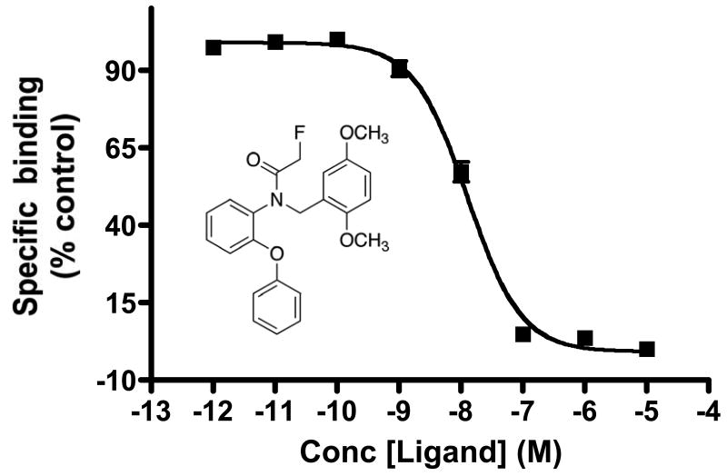
Translocator protein (TSPO), also referred to as peripheral benzodiazepine receptor (PBR), is a crucial 18-kDa outer mitochondrial membrane protein involved in numerous cellular functions, including the regulation of cholesterol metabolism, steroidogenesis, and apoptosis. Elevated expression of TSPO in oncology correlates with disease progression and poor survival, suggesting that molecular probes capable of assaying TSPO levels may have potential as cancer imaging biomarkers. In preclinical PET studies, we characterized a high-affinity aryloxyanilide-based TSPO imaging ligand, 18F-N-fluoroacetyl-N-(2,5-dimethoxybenzyl)-2-phenoxyaniline (18F-PBR06), as a candidate probe for the quantitative assessment of TSPO expression in glioma.
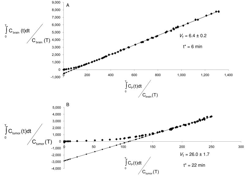
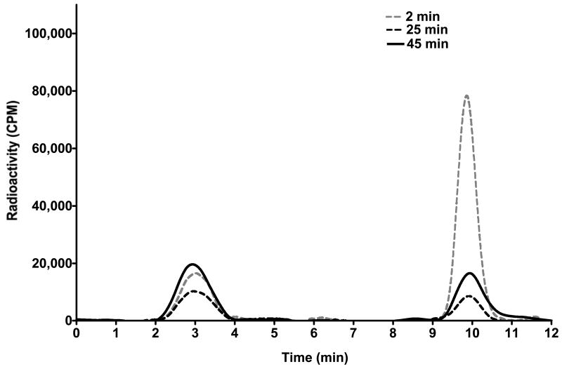
Glioma-bearing rats were imaged with 18F-PBR06 in a small-animal PET system. Dynamic images were acquired simultaneously on injection of 18F-PBR06 (70-100 MBq/0.2 mL). Over the course of scanning, arterial blood was collected to derive the input function, with high-performance liquid chromatography radiometabolite analysis performed on selected samples for arterial input function correction. Compartmental modeling of the PET data was performed using the corrected arterial input function. Specific tumor cell binding of PBR06 was evaluated by radioligand displacement of 3H-PK 11195 with PBR06 in vitro and by displacement of 18F-PBR06 with excess PBR06 in vivo. Immediately after imaging, tumor tissue and adjacent healthy brain were harvested for assay of TSPO protein levels by Western blotting and immunohistochemistry.
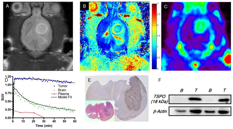
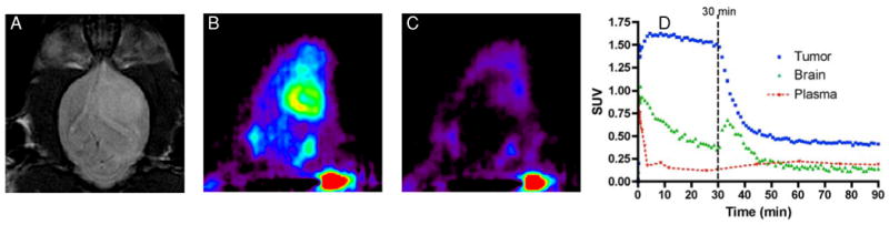
18F-PBR06 was found to preferentially accumulate in tumors, with modest uptake in the contralateral brain, facilitating excellent contrast between tumor and adjacent tissue. Infusion with PBR06 (10 mg/kg) displaced 18F-PBR06 binding by approximately 75%. The accumulation of 18F-PBR06 in tumor tissues and adjacent brain agreed with the ex vivo assay of TSPO protein levels by Western blotting and quantitative immunohistochemistry.
These preclinical studies illustrate that 18F-PBR06 is a promising tracer for visualization of TSPO-expressing tumors. Importantly, the close correlation between 18F-PBR06 uptake and TSPO expression in tumors and normal tissues, coupled with the high degree of displaceable binding from both tumors and the normal brain, represents a significant improvement over other TSPO imaging ligands previously evaluated in glioma. These data suggest the potential of 18F-PBR06 to elucidate the role of TSPO in oncology, as well as its potential development as a cancer imaging biomarker.
使用小动物 PET 系统对荷瘤大鼠进行 18F-PBR06 成像。在注射 18F-PBR06(70-100MBq/0.2mL)的同时,采集动态图像。在扫描过程中,采集动脉血以获得输入函数,并对选定的样本进行高效液相色谱放射性代谢产物分析,以进行动脉输入函数校正。使用校正后的动脉输入函数对 PET 数据进行房室模型分析。通过 3H-PK 11195 体外置换和体内过量 PBR06 置换评估 PBR06 的肿瘤细胞结合特异性。成像后,立即采集肿瘤组织和相邻正常脑组织,通过 Western blot 和免疫组化检测 TSPO 蛋白水平。
发现 18F-PBR06 优先在肿瘤中积聚,对侧大脑的摄取量适中,有利于肿瘤与相邻组织之间的良好对比。PBR06 输注(10mg/kg)置换 18F-PBR06 结合约 75%。18F-PBR06 在肿瘤组织和相邻脑组织中的积聚与 Western blot 和定量免疫组化检测 TSPO 蛋白水平的体外测定结果一致。
这些临床前研究表明,18F-PBR06 是一种有前途的 TSPO 表达肿瘤可视化示踪剂。重要的是,肿瘤和正常组织中 18F-PBR06 摄取与 TSPO 表达之间的密切相关性,以及肿瘤和正常脑组织中可置换结合的高度,与之前在神经胶质瘤中评估的其他 TSPO 成像配体相比有了显著的改进。这些数据表明 18F-PBR06 有潜力阐明 TSPO 在肿瘤学中的作用,以及作为癌症成像生物标志物的潜在发展。