Department of Microbiology, Escola Paulista de Medicina, Universidade Federal de São Paulo, São Paulo, Brazil.
PLoS Negl Trop Dis. 2010 Dec 7;4(12):e905. doi: 10.1371/journal.pntd.0000905.
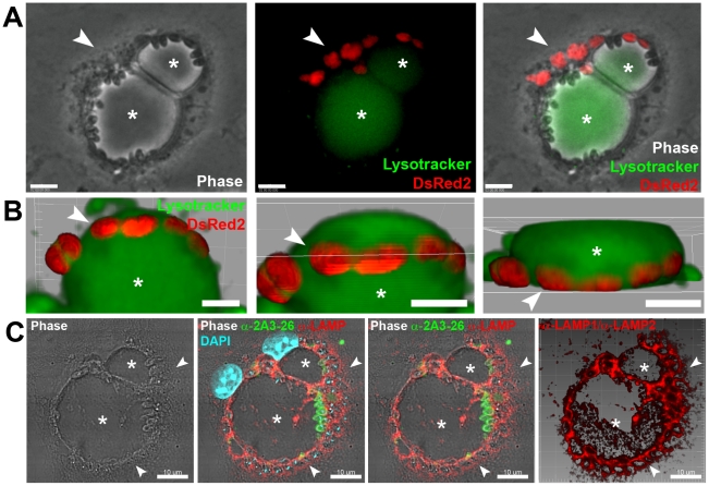
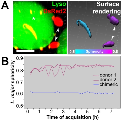
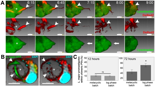
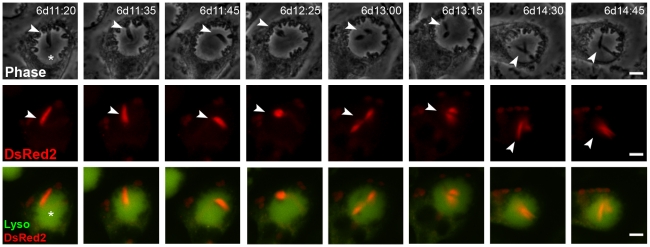
Protozoan parasites of the genus Leishmania alternate between flagellated, elongated extracellular promastigotes found in insect vectors, and round-shaped amastigotes enclosed in phagolysosome-like Parasitophorous Vacuoles (PVs) of infected mammalian host cells. Leishmania amazonensis amastigotes occupy large PVs which may contain many parasites; in contrast, single amastigotes of Leishmania major lodge in small, tight PVs, which undergo fission as parasites divide. To determine if PVs of these Leishmania species can fuse with each other, mouse macrophages in culture were infected with non-fluorescent L. amazonensis amastigotes and, 48 h later, superinfected with fluorescent L. major amastigotes or promastigotes. Fusion was investigated by time-lapse image acquisition of living cells and inferred from the colocalization of parasites of the two species in the same PVs. Survival, multiplication and differentiation of parasites that did or did not share the same vacuoles were also investigated. Fusion of PVs containing L. amazonensis and L. major amastigotes was not found. However, PVs containing L. major promastigotes did fuse with pre-established L. amazonensis PVs. In these chimeric vacuoles, L. major promastigotes remained motile and multiplied, but did not differentiate into amastigotes. In contrast, in doubly infected cells, within their own, unfused PVs metacyclic-enriched L. major promastigotes, but not log phase promastigotes--which were destroyed--differentiated into proliferating amastigotes. The results indicate that PVs, presumably customized by L. major amastigotes or promastigotes, differ in their ability to fuse with L. amazonensis PVs. Additionally, a species-specific PV was required for L. major destruction or differentiation--a requirement for which mechanisms remain unknown. The observations reported in this paper should be useful in further studies of the interactions between PVs to different species of Leishmania parasites, and of the mechanisms involved in the recognition and fusion of PVs.
原生动物寄生虫利什曼原虫属在有鞭毛的、拉长的细胞外前鞭毛体和被感染的哺乳动物宿主细胞的吞噬体样吞噬小体(PVs)内的圆形无鞭毛体阿米巴之间交替。利什曼原虫属亚马逊滋养体占据大的 PVs,其中可能含有许多寄生虫;相比之下,利什曼原虫属大滋养体的单个阿米巴虫栖息在小而紧密的 PVs 中,当寄生虫分裂时,PVs 会发生裂变。为了确定这些利什曼原虫物种的 PV 是否可以彼此融合,在培养的小鼠巨噬细胞中感染非荧光利什曼原虫属亚马逊滋养体,48 小时后,用荧光利什曼原虫属大滋养体或前鞭毛体再次感染。通过对活细胞的延时图像采集来研究融合,并根据两种物种的寄生虫在同一 PVs 中的共定位来推断融合。还研究了未共享同一空泡或共享同一空泡的寄生虫的存活、增殖和分化。未发现含有利什曼原虫属亚马逊滋养体和利什曼原虫属大滋养体的 PV 融合。然而,含有利什曼原虫属大前鞭毛体的 PV 确实与预先建立的利什曼原虫属亚马逊 PV 融合。在这些嵌合空泡中,利什曼原虫属大前鞭毛体保持运动和增殖,但不会分化为无鞭毛体。相比之下,在双重感染的细胞中,在它们自己的、未融合的 PVs 中,富含代谢型的利什曼原虫属大前鞭毛体,但不是对数期前鞭毛体——被破坏——分化为增殖的无鞭毛体。结果表明,PVs 可能由利什曼原虫属大前鞭毛体或无鞭毛体定制,在与利什曼原虫属亚马逊 PV 融合的能力上存在差异。此外,需要一种特定于物种的 PV 来破坏或分化利什曼原虫属大前鞭毛体——这一要求的机制尚不清楚。本文报道的观察结果应有助于进一步研究不同种利什曼原虫寄生虫之间的 PV 相互作用,以及参与 PV 识别和融合的机制。