Department of Animal Biology, Institute of Biology, State University of Campinas - UNICAMP, Campinas, Brazil.
Department of Biochemistry and Tissue Biology, Institute of Biology, State University of Campinas - UNICAMP, Campinas, Brazil.
Front Cell Infect Microbiol. 2022 Feb 4;12:824494. doi: 10.3389/fcimb.2022.824494. eCollection 2022.
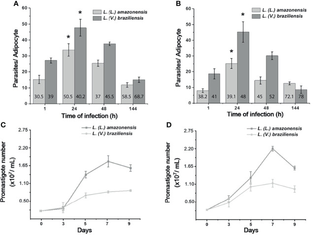
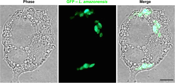
Although macrophages have long been considered key players in the course of infections, other non-professional phagocytes have lately been shown to maintain low levels of the parasite in safe intracellular niches. Recently, it was demonstrated that the adipose tissue is capable of harboring Old World () in mice. However, there is no evidence of experimental adipocyte infection with New World species so far. In addition, it was not known whether adipocytes would be permissive for formation of the unique, large and communal parasitophorous vacuoles that are typical of () in macrophages. Here we evaluated the ability of () and () promastigotes and amastigotes to infect 3T3-L1 fibroblast-derived adipocytes (3T3-Ad) using light and transmission electron microscopy. Our results indicate that amastigotes and promastigotes of both species were capable of infecting and surviving inside pre- and fully differentiated 3T3-Ad for up to 144 h. Importantly, () amastigotes resided in large communal parasitophorous vacuoles in pre-adipocytes, which appeared to be compressed between large lipid droplets in mature adipocytes. In parallel, individual amastigotes were detected in single vacuoles 144 h post-infection. We conclude that 3T3-Ad may constitute an environment that supports low loads of viable parasites perhaps contributing to parasite maintenance, since amastigotes of both species recovered from these cells differentiated into replicative promastigotes. Our findings shed light on the potential of a new host cell model that can be relevant to the persistence of New World species.
尽管巨噬细胞长期以来一直被认为是感染过程中的关键参与者,但最近其他非专业吞噬细胞也被证明能够在安全的细胞内龛位维持低水平的寄生虫。最近,研究表明脂肪组织能够在小鼠体内容纳旧世界()。然而,目前尚无实验性脂肪细胞感染新世界()物种的证据。此外,人们还不知道脂肪细胞是否允许形成独特的、大型的共寄生空泡,这种空泡是巨噬细胞中()的典型特征。在这里,我们使用光镜和透射电镜评估了()和()前体和无鞭毛体感染 3T3-L1 成纤维细胞衍生的脂肪细胞(3T3-Ad)的能力。我们的结果表明,两种物种的无鞭毛体和前体都能够感染和在预分化和完全分化的 3T3-Ad 内存活长达 144 小时。重要的是,()无鞭毛体在预脂肪细胞中存在于大型共寄生空泡中,这些空泡似乎在成熟脂肪细胞中的大脂质滴之间被压缩。同时,在感染后 144 小时,单个()无鞭毛体被检测到存在于单个空泡中。我们得出结论,3T3-Ad 可能构成一种支持低载量存活寄生虫的环境,这可能有助于寄生虫的维持,因为从这些细胞中回收的两种物种的无鞭毛体分化为复制性前体。我们的研究结果揭示了一种新的宿主细胞模型的潜力,该模型可能与新世界()物种的持续存在有关。