Department for Interface Science, Institute for Electronic Appliances and Circuits, University of Rostock, Albert-Einstein-Str. 2, 18059 Rostock, Germany.
Eur Biophys J. 2011 Mar;40(3):317-27. doi: 10.1007/s00249-010-0649-0. Epub 2010 Dec 14.
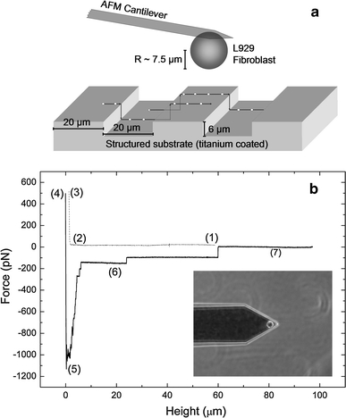
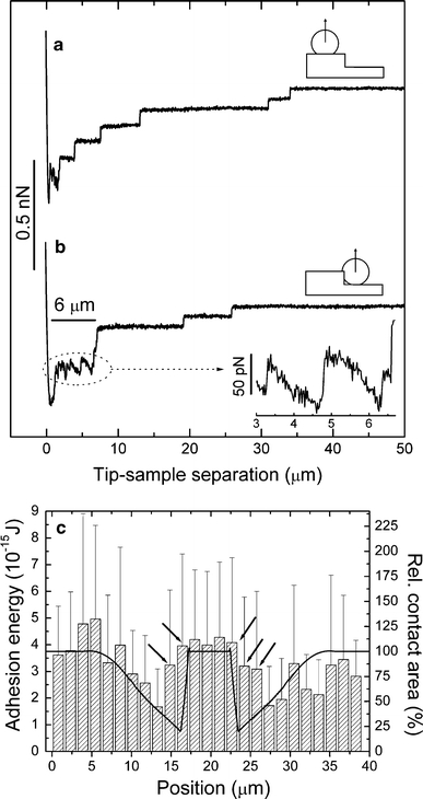
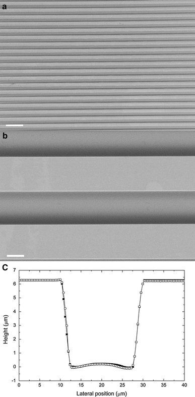
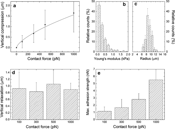
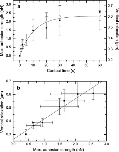
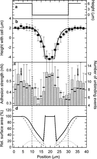
Single-cell force spectroscopy was used to investigate the initial adhesion of L929 fibroblasts onto periodically grooved titanium microstructures (height ~6 μm, groove width 20 μm). The position-dependent local adhesion strength of the cells was correlated with their rheological behavior. Spherical cells exhibited a significantly lower Young's modulus (<1 kPa) than that reported for spread cells, and their elastic properties can roughly be explained by the Hertz model for an elastic sphere. While in contact with the planar regions of the substrate, the cells started to adapt their shape through slight ventral flattening. The process was found to be independent of the applied contact force for values between 100 and 1,000 pN. The degree of flattening correlated with the adhesion strength during the first 60 s. Adhesion strength can be described by fast exponential kinetics as C₁[1-exp(-C₂·t] with C₁ = 2.34 ± 0.19 nN and C₂ = 0.09 ± 0.02 s⁻¹. A significant drop in the adhesion strength of up to 50% was found near the groove edges. The effect can be interpreted by the geometric decrease of the contact area, which indicates the inability of the fibroblasts to adapt to the shape of the substrate. Our results explain the role of the substrate's topography in contact guidance and suggest that rheological cell properties must be considered in cell adhesion modeling.
单细胞力谱技术被用于研究 L929 成纤维细胞最初在周期性沟槽钛微结构(高度约 6 μm,沟槽宽度 20 μm)上的黏附。细胞的位置相关局部黏附强度与它们的流变行为相关联。与铺展细胞相比,球形细胞表现出显著更低的杨氏模量(<1 kPa),并且它们的弹性性质可以通过赫兹模型对于弹性球体的解释来大致说明。当与基底的平面区域接触时,细胞开始通过轻微的腹侧变平来适应它们的形状。发现该过程与在 100 至 1000 pN 之间的施加接触力值无关。变平的程度与最初 60 秒内的黏附强度相关。黏附强度可以用快速指数动力学来描述,即 C₁[1-exp(-C₂·t)],其中 C₁ = 2.34 ± 0.19 nN,C₂ = 0.09 ± 0.02 s⁻¹。在沟槽边缘附近发现黏附强度显著下降了 50%。该效应可以通过接触面积的几何减小来解释,这表明成纤维细胞无法适应基底的形状。我们的结果解释了基底形貌在接触导向中的作用,并表明在细胞黏附建模中必须考虑流变细胞特性。