• 文献检索
  • 文档翻译
  • 深度研究
  • 学术资讯
  • Suppr Zotero 插件Zotero 插件
  • 邀请有礼
  • 套餐&价格
  • 历史记录
应用&插件
Suppr Zotero 插件Zotero 插件浏览器插件Mac 客户端Windows 客户端微信小程序
定价
高级版会员购买积分包购买API积分包
服务
文献检索文档翻译深度研究API 文档MCP 服务
关于我们
关于 Suppr公司介绍联系我们用户协议隐私条款
关注我们

Suppr 超能文献

核心技术专利:CN118964589B侵权必究
粤ICP备2023148730 号-1Suppr @ 2026

文献检索

告别复杂PubMed语法,用中文像聊天一样搜索,搜遍4000万医学文献。AI智能推荐,让科研检索更轻松。

立即免费搜索

文件翻译

保留排版,准确专业,支持PDF/Word/PPT等文件格式,支持 12+语言互译。

免费翻译文档

深度研究

AI帮你快速写综述,25分钟生成高质量综述,智能提取关键信息,辅助科研写作。

立即免费体验

组织对密封材料的反应:生物相容性的不同观点。

Tissue reaction to sealing materials: different view at biocompatibility.

机构信息

Institute of Pathology, REPAIR-Lab, Johannes Gutenberg University, Langenbeckstr. 1, 55101 Mainz, Germany.

出版信息

Eur J Med Res. 2010 Nov 25;15(11):483-92. doi: 10.1186/2047-783x-15-11-483.

DOI:10.1186/2047-783x-15-11-483
PMID:21159573
原文链接:https://pmc.ncbi.nlm.nih.gov/articles/PMC3352657/
Abstract

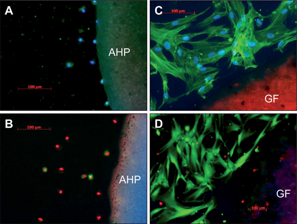
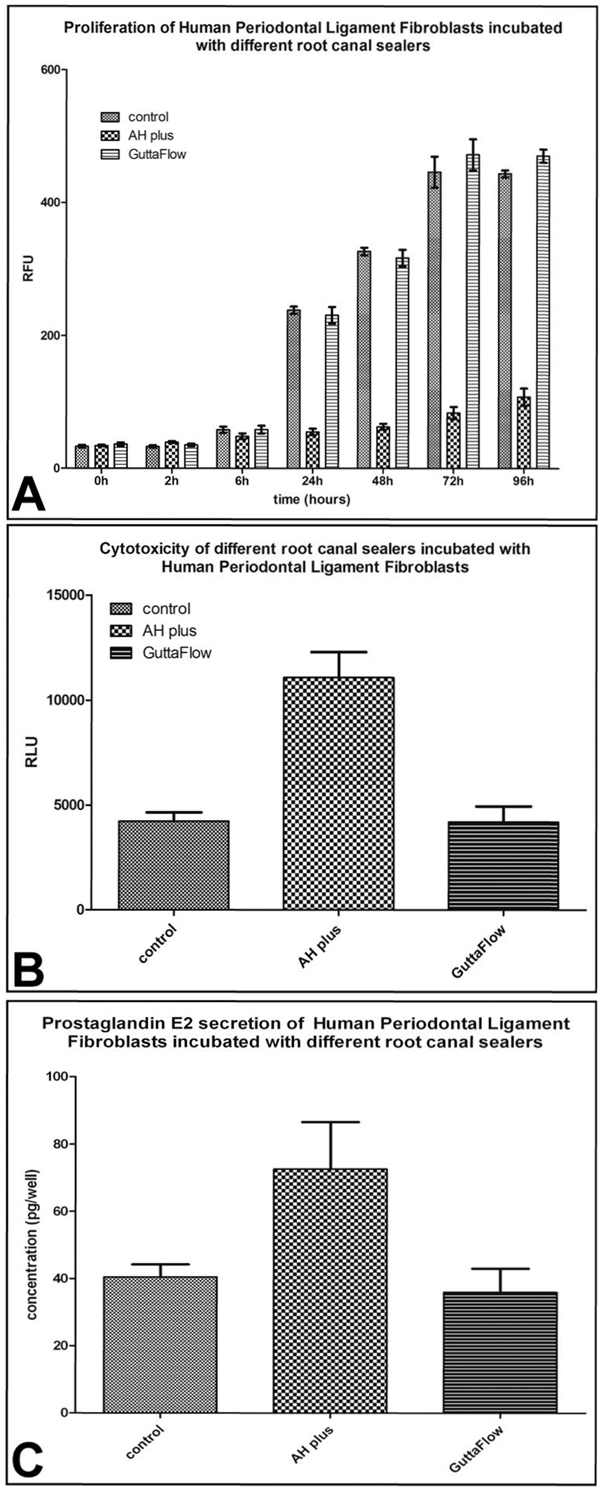
The biodegradability of root canal sealers in areas other than the root canal system is crucial to the overall success rate of endodontic treatment. The aim of the present study was to investigate, the cell and tissue reaction to GuttaFlow and AHPlus, both in vitro and in vivo. For the in vitro experiments the materials were incubated with Human Periodontal Ligament Fibroblasts and cell proliferation and cytotoxicity analyses were performed. Additional fluorescence-microscope stainings were carried out in order to visualize cell growth and morphology. For assessment of the tissue reaction to the materials a subcutaneous implantation model in Wistar rats was employed and the inflammatory response to the materials was visualized by means of general and specific histology after 6 weeks. Human gingival fibroblasts proliferation seemed to be dependent upon dental material and cultivation time. After an incubation period of 96 hrs AHPlus proved to be significantly (p<0.002) more cytotoxic than GuttaFlow, as only a small number of fibroblasts survived on AHPlus. In vivo, GuttaFlow was surrounded by a fibrous capsule and no degradation took place, while AHPlus induced a well-vascularized granulation tissue in which the material was phagocyted by macrophages. The results of this study demonstrate that a potential cytotoxic effect of a sealing material may beneficial in order to have antibacterial properties and induce self degradation when accidentally extruded over the apical foramen.

摘要

根管封闭剂在根管系统以外的区域的生物降解性对根管治疗的整体成功率至关重要。本研究的目的是研究 GuttaFlow 和 AHPlus 在体外和体内的细胞和组织反应。对于体外实验,将材料与人牙周膜成纤维细胞孵育,并进行细胞增殖和细胞毒性分析。此外,还进行了荧光显微镜染色,以观察细胞生长和形态。为了评估材料对组织的反应,采用了 Wistar 大鼠皮下植入模型,在 6 周后通过一般和特殊组织学观察材料的炎症反应。人牙龈成纤维细胞的增殖似乎取决于牙科材料和培养时间。在孵育 96 小时后,AHPlus 的细胞毒性明显(p<0.002)强于 GuttaFlow,因为只有少数成纤维细胞在 AHPlus 上存活。在体内,GuttaFlow 被纤维囊包围,没有发生降解,而 AHPlus 则诱导了富含血管的肉芽组织,其中巨噬细胞吞噬了材料。本研究的结果表明,密封材料的潜在细胞毒性作用可能有益于具有抗菌性能,并在意外挤出根尖孔上方时诱导自身降解。

https://cdn.ncbi.nlm.nih.gov/pmc/blobs/050f/3352657/a4f9b425e8cf/2047-783X-15-11-483-4.jpg
https://cdn.ncbi.nlm.nih.gov/pmc/blobs/050f/3352657/459bd97e901a/2047-783X-15-11-483-1.jpg
https://cdn.ncbi.nlm.nih.gov/pmc/blobs/050f/3352657/e69caf67c13a/2047-783X-15-11-483-2.jpg
https://cdn.ncbi.nlm.nih.gov/pmc/blobs/050f/3352657/dc94f184f6b5/2047-783X-15-11-483-3.jpg
https://cdn.ncbi.nlm.nih.gov/pmc/blobs/050f/3352657/a4f9b425e8cf/2047-783X-15-11-483-4.jpg

相似文献

1
Tissue reaction to sealing materials: different view at biocompatibility.组织对密封材料的反应:生物相容性的不同观点。
Eur J Med Res. 2010 Nov 25;15(11):483-92. doi: 10.1186/2047-783x-15-11-483.
2
A novel GuttaFlow sealer supports cell survival and attachment.一种新型的GuttaFlow封闭剂可支持细胞存活和附着。
J Endod. 2014 Feb;40(2):231-4. doi: 10.1016/j.joen.2013.08.029. Epub 2013 Oct 15.
3
[Effect of root canal sealers on biocompatibility of human periodontal ligament cells].[根管封闭剂对人牙周膜细胞生物相容性的影响]
Beijing Da Xue Xue Bao Yi Xue Ban. 2016 Oct 18;48(5):871-877.
4
A comparative study of the effects of three root-end filling materials on proliferation and adherence of human periodontal ligament fibroblasts.三种根充材料对人牙周膜成纤维细胞增殖及黏附影响的对比研究。
J Endod. 2011 Jun;37(6):865-70. doi: 10.1016/j.joen.2011.03.011.
5
Cytotoxicity of resin-, zinc oxide-eugenol-, and calcium hydroxide-based root canal sealers on human periodontal ligament cells and permanent V79 cells.树脂类、氧化锌丁香酚类和氢氧化钙类根管封闭剂对人牙周膜细胞和永生V79细胞的细胞毒性
Int Endod J. 2002 Feb;35(2):153-8. doi: 10.1046/j.1365-2591.2002.00459.x.
6
Cytotoxicity evaluation of various resin based root canal sealers.各种树脂基根管封闭剂的细胞毒性评价。
Int Endod J. 2010 Feb;43(2):148-53. doi: 10.1111/j.1365-2591.2009.01669.x.
7
Cytotoxicity of GuttaFlow Bioseal, GuttaFlow2, MTA Fillapex, and AH Plus on Human Periodontal Ligament Stem Cells.GuttaFlow Bioseal、GuttaFlow2、MTA Fillapex 和 AH Plus 对人牙周膜干细胞的细胞毒性。
J Endod. 2017 May;43(5):816-822. doi: 10.1016/j.joen.2017.01.001. Epub 2017 Mar 23.
8
Research that matters - biocompatibility and cytotoxicity screening.重要研究——生物相容性和细胞毒性筛选
Int Endod J. 2013 Mar;46(3):195-7. doi: 10.1111/iej.12047.
9
In vitro biocompatibility, inflammatory response, and osteogenic potential of 4 root canal sealers: Sealapex, Sankin apatite root sealer, MTA Fillapex, and iRoot SP root canal sealer.4种根管封闭剂的体外生物相容性、炎症反应和成骨潜力:Sealapex、三金磷灰石根管封闭剂、MTA Fillapex和iRoot SP根管封闭剂
J Endod. 2014 Oct;40(10):1642-8. doi: 10.1016/j.joen.2014.04.006. Epub 2014 Jun 7.
10
Effect of time of extraction on the biocompatibility of endodontic sealers with primary human fibroblasts.提取时间对原代人成纤维细胞与根管封闭剂生物相容性的影响。
Braz Oral Res. 2012 Sep-Oct;26(5):424-30. doi: 10.1590/s1806-83242012000500008.

引用本文的文献

1
Comparative analysis of the inflammatory response of human gingival fibroblasts to NeoSEALER Flo and CeraSeal bioceramic sealers: an in vitro study.人牙龈成纤维细胞对NeoSEALER Flo和CeraSeal生物陶瓷封闭剂炎症反应的比较分析:一项体外研究。
BMC Oral Health. 2025 Mar 17;25(1):395. doi: 10.1186/s12903-025-05692-1.
2
Analysis of Tissue Repair of a New Cement Based on Calcium and Strontium Aluminates: A Triple-Blinded, Randomized, Controlled Trial in an Animal Model.基于钙和锶铝酸盐的新型骨水泥组织修复分析:动物模型中的三盲、随机、对照试验
Eur J Dent. 2024 Oct;18(4):1076-1084. doi: 10.1055/s-0044-1786875. Epub 2024 May 22.
3

本文引用的文献

1
Histological and histomorphometrical analysis of a silica matrix embedded nanocrystalline hydroxyapatite bone substitute using the subcutaneous implantation model in Wistar rats.使用Wistar大鼠皮下植入模型对二氧化硅基质包埋的纳米晶羟基磷灰石骨替代物进行组织学和组织形态计量学分析。
Biomed Mater. 2010 Jun;5(3):35005. doi: 10.1088/1748-6041/5/3/035005. Epub 2010 May 11.
2
Evaluation of cytotoxicity of two root canal filling materials by MTT assay.通过MTT法评估两种根管充填材料的细胞毒性。
Aust Endod J. 2010 Apr;36(1):24-8. doi: 10.1111/j.1747-4477.2009.00173.x.
3
Fine-tuning scaffolds for tissue regeneration: effects of formic acid processing on tissue reaction to silk fibroin.
The Assessment of Quality of the Root Canal Filling and the Number of Visits Needed for Completing Primary Root Canal Treatment by Operators with Different Experience.
不同经验的操作者对根管充填质量及完成乳牙根管治疗所需就诊次数的评估
Bioengineering (Basel). 2022 Sep 13;9(9):468. doi: 10.3390/bioengineering9090468.
4
Experimental Study on the Cement-Based Materials Used in Coal Mine Gas Extraction for Hole Sealing.煤矿瓦斯抽采封孔用水泥基材料的试验研究
ACS Omega. 2021 Aug 3;6(32):21094-21103. doi: 10.1021/acsomega.1c02911. eCollection 2021 Aug 17.
5
Egyptian Propolis-Loaded Nanoparticles as a Root Canal Nanosealer: Sealing Ability and in vivo Biocompatibility.埃及蜂胶负载纳米粒子作为根管纳米密封剂:密封能力和体内生物相容性。
Int J Nanomedicine. 2020 Jul 27;15:5265-5277. doi: 10.2147/IJN.S258888. eCollection 2020.
6
Development of a novel histological and histomorphometric evaluation protocol for a standardized description of the mid-palatal suture - An ex vivo study.一种用于中隔缝标准化描述的新型组织学和组织形态计量学评估方案的制定 - 一项离体研究。
J Anat. 2019 Jul;235(1):180-188. doi: 10.1111/joa.12985. Epub 2019 Apr 4.
7
Effect of root canal sealers on human periodontal ligament fibroblast viability: ex vivo study.根管封闭剂对人牙周膜成纤维细胞活力的影响:体外研究
Odontology. 2018 Jul;106(3):245-256. doi: 10.1007/s10266-017-0329-y. Epub 2017 Dec 14.
8
A Highly Elastic and Rapidly Crosslinkable Elastin-Like Polypeptide-Based Hydrogel for Biomedical Applications.一种用于生物医学应用的基于高弹性且可快速交联的类弹性蛋白多肽的水凝胶。
Adv Funct Mater. 2015 Aug 12;25(30):4814-4826. doi: 10.1002/adfm.201501489. Epub 2015 Jul 1.
9
Biological responses of human gingival fibroblasts (HGFs) in an innovative co-culture model with Streptococcus mitis to thermosets coated with a silver polysaccharide antimicrobial system.人牙龈成纤维细胞(HGFs)在与缓症链球菌共培养的创新模型中对涂有银多糖抗菌系统的热固性材料的生物学反应。
PLoS One. 2014 May 7;9(5):e96520. doi: 10.1371/journal.pone.0096520. eCollection 2014.
10
In vivo biocompatibility of Resilon compared with gutta-percha in a pre-clinical model.在临床前模型中,Resilon与牙胶的体内生物相容性比较。
Dent Res J (Isfahan). 2013 Sep;10(5):652-8.
微调组织再生支架:甲酸处理对丝素蛋白组织反应的影响。
J Tissue Eng Regen Med. 2010 Aug;4(6):464-72. doi: 10.1002/term.257.
4
The induction of cytotoxicity, oxidative stress, and genotoxicity by root canal sealers in mammalian cells.根管封闭剂对哺乳动物细胞的细胞毒性、氧化应激及遗传毒性诱导作用
Oral Surg Oral Med Oral Pathol Oral Radiol Endod. 2009 Dec;108(6):952-60. doi: 10.1016/j.tripleo.2009.07.015.
5
Comparision of biocompatibility and cytotoxicity of two new root canal sealers.两种新型根管封闭剂的生物相容性和细胞毒性比较。
Acta Histochem. 2010 Nov;112(6):567-75. doi: 10.1016/j.acthis.2009.06.005. Epub 2009 Sep 22.
6
Dynamic in vivo biocompatibility of angiogenic peptide amphiphile nanofibers.血管生成肽两亲性纳米纤维的动态体内生物相容性
Biomaterials. 2009 Oct;30(31):6202-12. doi: 10.1016/j.biomaterials.2009.07.063. Epub 2009 Aug 15.
7
In vitro genotoxicity of root canal sealers.根管封闭剂的体外遗传毒性
Int Endod J. 2009 Mar;42(3):253-63. doi: 10.1111/j.1365-2591.2008.01510.x.
8
Evaluation of the biocompatibility of root canal sealers using subcutaneous implants.使用皮下植入物评估根管封闭剂的生物相容性。
J Appl Oral Sci. 2007 Jun;15(3):186-94. doi: 10.1590/s1678-77572007000300007.
9
Contribution of outgrowth endothelial cells from human peripheral blood on in vivo vascularization of bone tissue engineered constructs based on starch polycaprolactone scaffolds.人外周血来源的内皮细胞对基于淀粉聚己内酯支架的骨组织工程构建体体内血管化的贡献。
Biomaterials. 2009 Feb;30(4):526-34. doi: 10.1016/j.biomaterials.2008.09.058. Epub 2008 Oct 31.
10
Five-year follow-up of a root canal filling material in the maxillary sinus: a case report.上颌窦内一种根管充填材料的五年随访:病例报告
Oral Surg Oral Med Oral Pathol Oral Radiol Endod. 2008 Oct;106(4):e54-6. doi: 10.1016/j.tripleo.2008.06.008. Epub 2008 Aug 20.