Szczurko Grzegorz, Pawińska Małgorzata, Łuczaj-Cepowicz Elżbieta, Kierklo Anna, Marczuk-Kolada Grażyna, Hołownia Adam
Department of Integrated Dentistry, Medical University of Białystok, M. Skłodowska-Curie Street 24A, 15-276, Białystok, Poland.
Department of Pediatric Dentistry, Medical University of Białystok, J. Waszyngton Street 15A, 15-274, Białystok, Poland.
Odontology. 2018 Jul;106(3):245-256. doi: 10.1007/s10266-017-0329-y. Epub 2017 Dec 14.
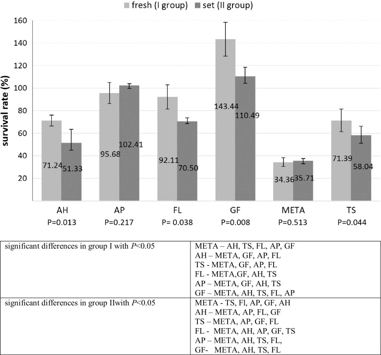
The aim of the study was to compare ex vivo the toxic effects of six root canal sealers immediately after mixing or setting on human periodontal ligament fibroblasts (HPdLF). Freshly mixed (I group) or set (allowed to dry for 24 h) (II group) specimens of AH Plus Jet (AH), Apexit Plus (AP), MTA Fillapex (FL), GuttaFlow (GF), MetaSEAL Soft (META), and Tubli-Seal (TS) were prepared. HPdLF were exposed for 24 h to the specimens. 3-(4,5-dimethylthiazolo-2-yl)-2,5-diphenyltetrazolium bromide assay was used to examine the effect of the root canal sealers on mitochondrial metabolic activity. Fluorescein isothiocyanate (FITC)-annexin V (AnV) and propidium iodide staining followed by flow cytometry was used to identify the effects of the materials on cell apoptosis/necrosis. Statistical analyses were performed by one-way ANOVA followed by post hoc tests, and significance was determined at P < 0.05. Most materials from the two groups reduced the viability of the cultured cells compared with the control group (P < 0.05). Statistical analysis showed significant differences in HPdLF viability between the individual materials in each group (P < 0.001). AH and AP induced a significant increase in the percentage of apoptotic cells, while TS, FL, and META elevated the proportion of necrotic cells compared with other materials and the controls (p < 0.05). The cytotoxic effects of the tested root canal sealers (both fresh and set) on HPdLF varied. Both forms of sealers were able to cause toxic effects by inducing apoptosis and necrosis in HPdLF. The cytotoxicity of FL, META, TS was mainly associated with necrosis, while AH and AP with apoptosis.
本研究的目的是在体外比较六种根管封闭剂在混合或凝固后立即对人牙周膜成纤维细胞(HPdLF)的毒性作用。制备了AH Plus Jet(AH)、Apexit Plus(AP)、MTA Fillapex(FL)、GuttaFlow(GF)、MetaSEAL Soft(META)和Tubli-Seal(TS)的新鲜混合样本(I组)或凝固样本(晾干24小时)(II组)。将HPdLF暴露于这些样本24小时。采用3-(4,5-二甲基噻唑-2-基)-2,5-二苯基四氮唑溴盐法检测根管封闭剂对线粒体代谢活性的影响。采用异硫氰酸荧光素(FITC)-膜联蛋白V(AnV)和碘化丙啶染色,随后进行流式细胞术,以确定材料对细胞凋亡/坏死的影响。采用单因素方差分析及事后检验进行统计分析,以P<0.05为差异有统计学意义。与对照组相比,两组中的大多数材料均降低了培养细胞的活力(P<0.05)。统计分析表明,每组中各材料之间HPdLF活力存在显著差异(P<0.001)。与其他材料及对照组相比,AH和AP诱导凋亡细胞百分比显著增加,而TS、FL和META则使坏死细胞比例升高(P<0.05)。测试的根管封闭剂(新鲜和凝固的)对HPdLF的细胞毒性作用各不相同。两种形式的封闭剂均能够通过诱导HPdLF凋亡和坏死而产生毒性作用。FL、META、TS的细胞毒性主要与坏死相关,而AH和AP的细胞毒性主要与凋亡相关。