CNRS UPR9021, Institut de Biologie Moléculaire et Cellulaire, 67000 Strasbourg, France.
Ann Rheum Dis. 2011 May;70(5):837-43. doi: 10.1136/ard.2010.139832. Epub 2010 Dec 20.
The P140 phosphopeptide issued from the spliceosomal U1-70K small nuclear ribonucleoprotein protein displays protective properties in MRL/lpr lupus-prone mice. It binds both major histocompatibility class II (MHCII) and HSC70/Hsp73 molecules. P140 peptide increases MRL/lpr peripheral blood lymphocyte apoptosis and decreases autoepitope recognition by T cells.
To explore further the mode of action of P140 peptide on HSC70+ antigen-presenting cells.
P140 biodistribution was monitored in real time using an imaging system and by fluorescence and electron microscopy. Fluorescence activated cell sorting and Western blotting experiments were used to evaluate the P140 effects on autophagic flux markers.
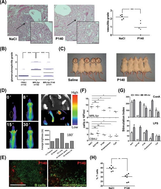
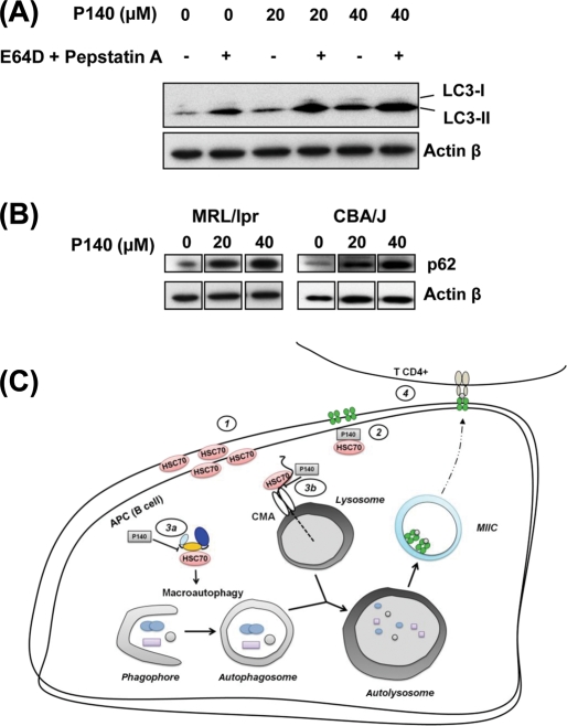
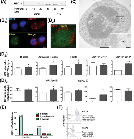
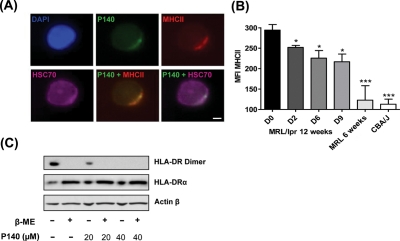
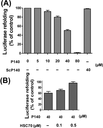
P140 fluorescence accumulated especially in the lungs and spleen. P140 peptide reduced the number of peripheral and splenic T and B cells without affecting these cells in normal mice. Remaining MRL/lpr B cells responded normally to mitogens. P140 peptide decreased the expression levels of HSC70/Hsp73 chaperone and stable MHCII dimers, which are both increased in MRL/lpr splenic B cells. It impaired refolding properties of chaperone HSC70. In MRL/lpr B cells, it increased the accumulation of the autophagy markers p62/SQSTM1 and LC3-II, consistent with a downregulated lysosomal degradation during autophagic flux.
The study results suggest that after P140 peptide binding to HSC70, the endogenous (auto)antigen processing might be greatly affected in MRL/lpr antigen-presenting B cells, leading to the observed decrease of autoreactive T-cell priming and signalling via a mechanism involving a lysosomal degradation pathway. This unexpected mechanism might explain the beneficial effect of P140 peptide in treated MRL/lpr mice.
从剪接体 U1-70K 小核核糖核蛋白蛋白中释放的 P140 磷酸肽在 MRL/lpr 狼疮易感小鼠中显示出保护特性。它与主要组织相容性复合体 II(MHCII)和 HSC70/Hsp73 分子都有结合。P140 肽增加 MRL/lpr 外周血淋巴细胞凋亡,并减少 T 细胞对自身表位的识别。
进一步探索 P140 肽对 HSC70+抗原呈递细胞的作用方式。
使用成像系统和荧光及电子显微镜实时监测 P140 的分布。使用荧光激活细胞分选和 Western blot 实验评估 P140 对自噬流标志物的影响。
P140 荧光在肺部和脾脏中特别积聚。P140 肽减少外周和脾脏 T 和 B 细胞的数量,但对正常小鼠的这些细胞没有影响。残留的 MRL/lpr B 细胞对有丝分裂原的反应正常。P140 肽降低了 HSC70/Hsp73 伴侣和稳定 MHCII 二聚体的表达水平,这些在 MRL/lpr 脾脏 B 细胞中都增加了。它损害了伴侣 HSC70 的重折叠特性。在 MRL/lpr B 细胞中,它增加了自噬标记物 p62/SQSTM1 和 LC3-II 的积累,与自噬流中溶酶体降解的下调一致。
研究结果表明,P140 肽与 HSC70 结合后,MRL/lpr 抗原呈递 B 细胞中的内源性(自身)抗原加工可能受到很大影响,导致观察到的自身反应性 T 细胞启动和信号转导减少,其机制涉及溶酶体降解途径。这种意外的机制可能解释了 P140 肽在治疗 MRL/lpr 小鼠中的有益作用。