Valenti Denise A
Vision Care, 62 Forest Avenue Quincy, MA 02169, USA.
Int J Alzheimers Dis. 2011 Jan 5;2010:793931. doi: 10.4061/2010/793931.
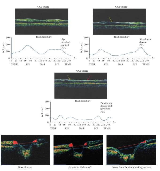
Imaging through the visual system in Alzheimer's disease, with the technology currently in widespread use for the diagnosis and management of eye disease such as glaucoma and macular degeneration, is proving to be promising. In vivo cross-section imaging during an annual comprehensive eye exam has been available for a decade for glaucoma and macular degeneration, and this same imaging, using Optical Coherence Tomography, has been demonstrated to show deficits specific to AD and mild cognitive impairment. These deficits are in the form of nerve fiber layer tissue drop out in the retina and optic nerve. The retrograde loss of nerve fiber layer tissue in the retina and optic nerve may be an early biomarker of AD, and these deficits in the nerve fiber layer of the retina and optic nerve may be the earliest sign of AD, even prior to damage to the hippocampal region that impacts memory.
利用目前广泛应用于青光眼和黄斑变性等眼部疾病诊断和管理的技术,对阿尔茨海默病患者的视觉系统进行成像,已被证明具有前景。在年度全面眼科检查期间进行的体内横断面成像,用于青光眼和黄斑变性已有十年,而使用光学相干断层扫描的这种相同成像已被证明能显示出阿尔茨海默病和轻度认知障碍特有的缺陷。这些缺陷表现为视网膜和视神经中神经纤维层组织缺失。视网膜和视神经中神经纤维层组织的逆行性丧失可能是阿尔茨海默病的早期生物标志物,而视网膜和视神经神经纤维层的这些缺陷可能是阿尔茨海默病的最早迹象,甚至早于影响记忆的海马区受损之前。