• 文献检索
  • 文档翻译
  • 深度研究
  • 学术资讯
  • Suppr Zotero 插件Zotero 插件
  • 邀请有礼
  • 套餐&价格
  • 历史记录
应用&插件
Suppr Zotero 插件Zotero 插件浏览器插件Mac 客户端Windows 客户端微信小程序
定价
高级版会员购买积分包购买API积分包
服务
文献检索文档翻译深度研究API 文档MCP 服务
关于我们
关于 Suppr公司介绍联系我们用户协议隐私条款
关注我们

Suppr 超能文献

核心技术专利:CN118964589B侵权必究
粤ICP备2023148730 号-1Suppr @ 2026

文献检索

告别复杂PubMed语法,用中文像聊天一样搜索,搜遍4000万医学文献。AI智能推荐,让科研检索更轻松。

立即免费搜索

文件翻译

保留排版,准确专业,支持PDF/Word/PPT等文件格式,支持 12+语言互译。

免费翻译文档

深度研究

AI帮你快速写综述,25分钟生成高质量综述,智能提取关键信息,辅助科研写作。

立即免费体验

光学相干断层扫描在非黑素细胞性皮肤肿瘤成像中的临床应用:一项多模态初步研究。

Clinical application of optical coherence tomography for the imaging of non-melanocytic cutaneous tumors: a pilot multi-modal study.

作者信息

Forsea Ana-Maria, Carstea Elfrieda Mihaela, Ghervase Luminita, Giurcaneanu Calin, Pavelescu Gabriela

机构信息

Dermatology Department, Elias University Hospital, Bucharest, Romania.

出版信息

J Med Life. 2010 Oct-Dec;3(4):381-9.

PMID:21254735
原文链接:https://pmc.ncbi.nlm.nih.gov/articles/PMC3019059/
Abstract

CONTEXT

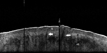
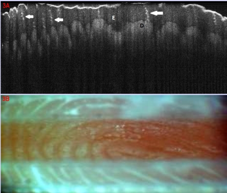
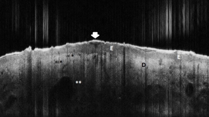
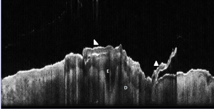
Optical coherence tomography (OCT) is an emergent imaging technique, based on the interference of infrared radiation and living tissues, that allows the in vivo visualization of the skin structures, at high resolution and up to 1.6 mm depth. As such, there is mounting evidence that OCT may be an interesting technique for the diagnosis of skin diseases, including the noninvasive early detection of cutaneous tumors.

OBJECTIVE

We aimed to investigate the utility of OCT for the diagnosis of non-melanocytic, non-pigmented cutaneous tumors.

METHODS

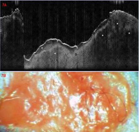
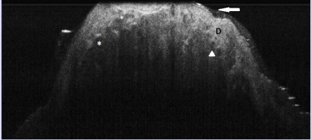
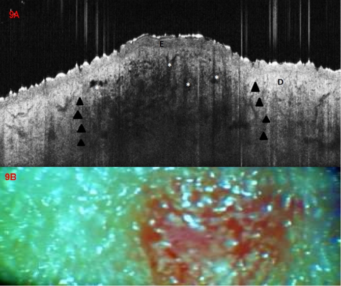
Preliminary results are presented from an initiated study. Fifteen consecutive patients with clinical suspicion of epithelial cancers and precancers registered over one week in a university dermatologic department were included. As control were selected 7 patients with inflammatory skin diseases (psoriasis, lichen planus, cutaneous lupus erythematosus). In all study and control patients, the lesions and samples of normal, perilesional skin were documented by clinical digital photography, contact dermoscopy with digital image capture and OCT with central wavelength of 930 nm. Final diagnosis was certified by histopathological analysis.

RESULTS

We could identify morphological features in OCT examination that distinguished between normal and lesional skin, and between neoplastic vs. inflammatory lesions. In the same time, combining OCT and dermatoscopical evaluation of a lesion improved the performance of diagnosis when compared to clinical diagnosis alone and with either OCT or dermoscopy imaging used alone.

CONCLUSIONS

OCT appears as a promising method of in vivo diagnosis of early neoplastic cutaneous lesions with equivocal clinical and/or dermoscopic aspect. Continuation of our study as well as other larger investigation will be able to contribute with new insights in the role of OCT in the non-invasive diagnosis of skin disease.

摘要

背景

光学相干断层扫描(OCT)是一种新兴的成像技术,基于红外辐射与活体组织的干涉原理,能够在体内以高分辨率对皮肤结构进行可视化成像,深度可达1.6毫米。因此,越来越多的证据表明,OCT可能是一种用于诊断皮肤疾病的有趣技术,包括皮肤肿瘤的无创早期检测。

目的

我们旨在研究OCT在诊断非黑素细胞性、无色素性皮肤肿瘤中的应用价值。

方法

本文呈现了一项已启动研究的初步结果。纳入了在一所大学皮肤科连续登记的15例临床怀疑为上皮癌和癌前病变的患者,这些患者在一周内就诊。选择7例患有炎症性皮肤病(银屑病、扁平苔藓、皮肤红斑狼疮)的患者作为对照。对所有研究对象和对照患者的病变以及正常、病变周围皮肤样本,通过临床数码摄影、带数字图像采集的接触式皮肤镜检查以及中心波长为930纳米的OCT进行记录。最终诊断通过组织病理学分析得以证实。

结果

我们能够在OCT检查中识别出区分正常皮肤与病变皮肤、肿瘤性病变与炎症性病变的形态学特征。同时,与单独的临床诊断以及单独使用OCT或皮肤镜成像相比,联合使用OCT和皮肤镜对病变进行评估可提高诊断性能。

结论

对于临床和/或皮肤镜表现不明确的早期皮肤肿瘤性病变,OCT似乎是一种有前景的体内诊断方法。继续开展我们的研究以及其他更大规模的调查,将能够为OCT在皮肤疾病无创诊断中的作用提供新的见解。

https://cdn.ncbi.nlm.nih.gov/pmc/blobs/fd9d/3019059/94faa318e044/JMedLife-03-381-g0012.jpg
https://cdn.ncbi.nlm.nih.gov/pmc/blobs/fd9d/3019059/98f4a1b66c6d/JMedLife-03-381-g001.jpg
https://cdn.ncbi.nlm.nih.gov/pmc/blobs/fd9d/3019059/4a1930d46b9f/JMedLife-03-381-g002.jpg
https://cdn.ncbi.nlm.nih.gov/pmc/blobs/fd9d/3019059/c3c4ad7b1674/JMedLife-03-381-g003.jpg
https://cdn.ncbi.nlm.nih.gov/pmc/blobs/fd9d/3019059/a12ccf79418d/JMedLife-03-381-g004.jpg
https://cdn.ncbi.nlm.nih.gov/pmc/blobs/fd9d/3019059/0a031b4927ac/JMedLife-03-381-g005.jpg
https://cdn.ncbi.nlm.nih.gov/pmc/blobs/fd9d/3019059/17d6558ea6a6/JMedLife-03-381-g006.jpg
https://cdn.ncbi.nlm.nih.gov/pmc/blobs/fd9d/3019059/d897c81fa1ea/JMedLife-03-381-g007.jpg
https://cdn.ncbi.nlm.nih.gov/pmc/blobs/fd9d/3019059/22263a8f9cce/JMedLife-03-381-g008.jpg
https://cdn.ncbi.nlm.nih.gov/pmc/blobs/fd9d/3019059/4bd8f0a2b3d5/JMedLife-03-381-g009.jpg
https://cdn.ncbi.nlm.nih.gov/pmc/blobs/fd9d/3019059/88b1e901f806/JMedLife-03-381-g0010.jpg
https://cdn.ncbi.nlm.nih.gov/pmc/blobs/fd9d/3019059/419dc55960b3/JMedLife-03-381-g0011.jpg
https://cdn.ncbi.nlm.nih.gov/pmc/blobs/fd9d/3019059/94faa318e044/JMedLife-03-381-g0012.jpg

相似文献

1
Clinical application of optical coherence tomography for the imaging of non-melanocytic cutaneous tumors: a pilot multi-modal study.光学相干断层扫描在非黑素细胞性皮肤肿瘤成像中的临床应用:一项多模态初步研究。
J Med Life. 2010 Oct-Dec;3(4):381-9.
2
In vivo microvascular imaging of cutaneous actinic keratosis, Bowen's disease and squamous cell carcinoma using dynamic optical coherence tomography.应用动态光学相干断层扫描对光化性角化病、鲍恩病和鳞状细胞癌的皮肤微血管进行体内成像。
J Eur Acad Dermatol Venereol. 2017 Oct;31(10):1655-1662. doi: 10.1111/jdv.14335. Epub 2017 Jun 20.
3
Role of In Vivo Reflectance Confocal Microscopy in the Analysis of Melanocytic Lesions.体内反射共聚焦显微镜在黑素细胞性病变分析中的作用
Acta Dermatovenerol Croat. 2018 Apr;26(1):64-67.
4
Application of Cellular Resolution Full-Field Optical Coherence Tomography in vivo for the Diagnosis of Skin Tumours and Inflammatory Skin Diseases: A Pilot Study.细胞分辨率全场光学相干断层成像在体内诊断皮肤肿瘤和炎症性皮肤病中的应用:一项初步研究。
Dermatology. 2022;238(1):121-131. doi: 10.1159/000514686. Epub 2021 May 4.
5
Diagnostic accuracy of optical coherence tomography in actinic keratosis and basal cell carcinoma.光学相干断层扫描在光化性角化病和基底细胞癌中的诊断准确性
Photodiagnosis Photodyn Ther. 2016 Dec;16:44-49. doi: 10.1016/j.pdpdt.2016.08.004. Epub 2016 Aug 9.
6
Line-field confocal optical coherence tomography in melanocytic and non-melanocytic skin tumors.线阵共焦光学相干断层扫描在黑素细胞和非黑素细胞性皮肤肿瘤中的应用。
Ital J Dermatol Venerol. 2023 Jun;158(3):180-189. doi: 10.23736/S2784-8671.23.07639-9.
7
Optical coherence tomography imaging of non-melanoma skin cancer undergoing imiquimod therapy.光相干断层扫描成像在咪喹莫特治疗非黑素瘤皮肤癌中的应用。
Skin Res Technol. 2014 May;20(2):170-6. doi: 10.1111/srt.12102. Epub 2013 Sep 16.
8
Optical coherence tomography and its role for delineating the thickness of keratinocyte dysplasia and neoplasia.光学相干断层扫描及其在描绘角质形成细胞发育异常和肿瘤厚度方面的作用。
Curr Probl Dermatol. 2015;46:95-100. doi: 10.1159/000366543. Epub 2014 Dec 18.
9
Comparison of different optical coherence tomography devices for diagnosis of non-melanoma skin cancer.不同光学相干断层扫描设备用于诊断非黑色素瘤皮肤癌的比较。
Skin Res Technol. 2016 Nov;22(4):395-405. doi: 10.1111/srt.12277. Epub 2016 Jan 25.
10
Optical Coherence Tomography in the Diagnosis of Skin Cancer.光学相干断层扫描在皮肤癌诊断中的应用。
Dermatol Clin. 2017 Oct;35(4):465-488. doi: 10.1016/j.det.2017.06.008. Epub 2017 Aug 9.

引用本文的文献

1
Interobserver agreement on line-field confocal optical coherence tomography image markers in keratinocyte carcinomas and precursor lesions.角化细胞癌及其前体病变中线扫描共焦光学相干断层成像图像标志物的观察者间一致性。
Arch Dermatol Res. 2024 Sep 6;316(8):608. doi: 10.1007/s00403-024-03344-y.
2
An Update on Non-Invasive Skin Imaging Techniques in Actinic Keratosis-A Narrative Review.光化性角化病的无创性皮肤成像技术更新:综述
Medicina (Kaunas). 2024 Jun 26;60(7):1043. doi: 10.3390/medicina60071043.
3
Line-Field Confocal Optical Coherence Tomography for the Diagnosis of Skin Tumors: A Systematic Review and Meta-Analysis.

本文引用的文献

1
Noninvasive imaging of living human skin with dual-wavelength optical coherence tomography in two and three dimensions.采用双波长光学相干断层扫描技术对活体人类皮肤进行二维和三维无创成像。
J Biomed Opt. 1998 Oct;3(4):446-55. doi: 10.1117/1.429897.
2
Simultaneous dual-band optical coherence tomography in the spectral domain for high resolution in vivo imaging.用于高分辨率体内成像的光谱域同步双波段光学相干断层扫描技术。
Opt Express. 2009 Oct 26;17(22):19486-500. doi: 10.1364/OE.17.019486.
3
Optical coherence tomography imaging of psoriasis vulgaris: correlation with histology and disease severity.
用于皮肤肿瘤诊断的线场共聚焦光学相干断层扫描:一项系统评价和荟萃分析
Diagnostics (Basel). 2024 Jul 15;14(14):1522. doi: 10.3390/diagnostics14141522.
4
Optical Coherence Tomography as a Valuable Tool for the Evaluation of Cutaneous Kaposi Sarcoma Treated with Imiquimod 5% Cream.光学相干断层扫描作为评估5%咪喹莫特乳膏治疗皮肤卡波西肉瘤的重要工具
Diagnostics (Basel). 2023 Sep 11;13(18):2901. doi: 10.3390/diagnostics13182901.
5
Role of modern imaging techniques for the diagnosis of lichen planus.现代成像技术在扁平苔藓诊断中的作用
Exp Ther Med. 2019 Feb;17(2):1052-1060. doi: 10.3892/etm.2018.6974. Epub 2018 Nov 16.
6
Optical coherence tomography for diagnosing skin cancer in adults.光学相干断层扫描技术在成人皮肤癌诊断中的应用
Cochrane Database Syst Rev. 2018 Dec 4;12(12):CD013189. doi: 10.1002/14651858.CD013189.
7
The clinical usefulness of optical coherence tomography during cancer interventions.光学相干断层扫描在癌症介入治疗中的临床应用。
J Cancer Res Clin Oncol. 2018 Oct;144(10):1967-1990. doi: 10.1007/s00432-018-2690-9. Epub 2018 Jun 20.
8
Noninvasive Long-term Monitoring of Actinic Keratosis and Field Cancerization Following Treatment with Ingenol Mebutate Gel 0.015.使用0.015% 鬼臼毒素凝胶治疗后光化性角化病和场癌化的无创长期监测
J Clin Aesthet Dermatol. 2017 Oct;10(10):28-33. Epub 2017 Oct 1.
9
Combined reflectance confocal microscopy-optical coherence tomography for delineation of basal cell carcinoma margins: an ex vivo study.联合反射共聚焦显微镜-光学相干断层扫描用于确定基底细胞癌边缘:一项离体研究。
J Biomed Opt. 2016 Jan;21(1):16006. doi: 10.1117/1.JBO.21.1.016006.
10
Motion correction of in vivo three-dimensional optical coherence tomography of human skin using a fiducial marker.使用基准标记对人体皮肤的体内三维光学相干断层扫描进行运动校正。
Biomed Opt Express. 2012 Aug 1;3(8):1774-86. doi: 10.1364/BOE.3.001774. Epub 2012 Jun 29.
光学相干断层扫描成像在寻常型银屑病中的应用:与组织病理学和疾病严重程度的相关性。
Arch Dermatol Res. 2010 Mar;302(2):105-11. doi: 10.1007/s00403-009-1000-4. Epub 2009 Nov 6.
4
Optical coherence tomography for imaging of skin and skin diseases.用于皮肤及皮肤疾病成像的光学相干断层扫描技术
Semin Cutan Med Surg. 2009 Sep;28(3):196-202. doi: 10.1016/j.sder.2009.07.002.
5
Angle-resolved optical coherence tomography with sequential angular selectivity for speckle reduction.具有顺序角度选择性以减少散斑的角度分辨光学相干断层扫描技术。
Opt Express. 2007 May 14;15(10):6200-9. doi: 10.1364/oe.15.006200.
6
Assessment of optical coherence tomography imaging in the diagnosis of non-melanoma skin cancer and benign lesions versus normal skin: observer-blinded evaluation by dermatologists and pathologists.光学相干断层扫描成像在非黑色素瘤皮肤癌及良性病变与正常皮肤诊断中的评估:皮肤科医生和病理学家的观察者盲法评估
Dermatol Surg. 2009 Jun;35(6):965-72. doi: 10.1111/j.1524-4725.2009.01164.x. Epub 2009 Apr 8.
7
Machine-learning classification of non-melanoma skin cancers from image features obtained by optical coherence tomography.基于光学相干断层扫描获得的图像特征对非黑色素瘤皮肤癌进行机器学习分类。
Skin Res Technol. 2008 Aug;14(3):364-9. doi: 10.1111/j.1600-0846.2008.00304.x.
8
Morphology and epidermal thickness of normal skin imaged by optical coherence tomography.通过光学相干断层扫描成像的正常皮肤的形态学和表皮厚度。
Dermatology. 2008;217(1):14-20. doi: 10.1159/000118508. Epub 2008 Feb 29.
9
Application of optical coherence tomography in non-invasive characterization of skin vascular lesions.光学相干断层扫描在皮肤血管病变无创特征分析中的应用。
Skin Res Technol. 2008 Feb;14(1):89-92. doi: 10.1111/j.1600-0846.2007.00265.x.
10
Using optical coherence tomography to evaluate skin sun damage and precancer.使用光学相干断层扫描技术评估皮肤晒伤和癌前病变。
Lasers Surg Med. 2007 Oct;39(9):687-95. doi: 10.1002/lsm.20573.