• 文献检索
  • 文档翻译
  • 深度研究
  • 学术资讯
  • Suppr Zotero 插件Zotero 插件
  • 邀请有礼
  • 套餐&价格
  • 历史记录
应用&插件
Suppr Zotero 插件Zotero 插件浏览器插件Mac 客户端Windows 客户端微信小程序
定价
高级版会员购买积分包购买API积分包
服务
文献检索文档翻译深度研究API 文档MCP 服务
关于我们
关于 Suppr公司介绍联系我们用户协议隐私条款
关注我们

Suppr 超能文献

核心技术专利:CN118964589B侵权必究
粤ICP备2023148730 号-1Suppr @ 2026

文献检索

告别复杂PubMed语法,用中文像聊天一样搜索,搜遍4000万医学文献。AI智能推荐,让科研检索更轻松。

立即免费搜索

文件翻译

保留排版,准确专业,支持PDF/Word/PPT等文件格式,支持 12+语言互译。

免费翻译文档

深度研究

AI帮你快速写综述,25分钟生成高质量综述,智能提取关键信息,辅助科研写作。

立即免费体验

视网膜静脉阻塞:视网膜静脉调节异常的潜在影响。

Retinal vein occlusions: The potential impact of a dysregulation of the retinal veins.

作者信息

Fraenkl Stephan A, Mozaffarieh Maneli, Flammer Josef

机构信息

Department of Ophthalmology, University of Basel, Mittlere Strasse 91, 4031 Basel, Switzerland.

出版信息

EPMA J. 2010 Jun;1(2):253-261. doi: 10.1007/s13167-010-0025-2. Epub 2010 Jun 18.

DOI:10.1007/s13167-010-0025-2
PMID:21258633
原文链接:https://pmc.ncbi.nlm.nih.gov/articles/PMC3003793/
Abstract

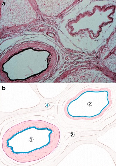
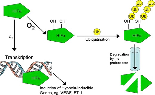
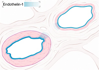
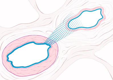
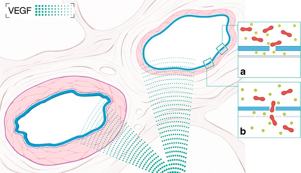
A retinal vein occlusion (RVO) is a sight threatening disease. It can be divided into central vein occlusion and branch retinal vein occlusion. The pathogenesis of the condition remains to be solved. Mechanical compression of the vessel wall or thrombotic occlusion of the vessel lumen, sometimes combined with rheological disorders, are often assumed pathomechanisms. Accordingly, the therapy relies either on mechanical decompression, lyses of thrombi or improvement of rheology. A number of observations however, such as the relationship of RVO to atherosclerotic risk factors, spontaneous reversibility particularly in young patients, rest flow observed in angiography, occlusion despite anticoagulation or thrombocytopenia and finally the positive effect of anti-VEGF therapy are not explained by the present pathogenetic concept. As a new concept we propose a local venous constriction induced by vasoconstrictive molecules diffusing from neighbouring diseased arteries and/or from other neighbouring (hypoxic) tissues. Recognizing these postulated conditions might lead to an earlier identification of impending vein occlusions as well as to a treatment more tailored to the risk factor constellation of the particular patient.

摘要

视网膜静脉阻塞(RVO)是一种威胁视力的疾病。它可分为中央静脉阻塞和视网膜分支静脉阻塞。该病的发病机制仍有待解决。血管壁的机械性压迫或血管腔的血栓性阻塞,有时伴有流变学紊乱,常被认为是发病机制。因此,治疗要么依赖于机械减压、血栓溶解或流变学改善。然而,一些观察结果,如RVO与动脉粥样硬化危险因素的关系、特别是年轻患者的自发可逆性、血管造影中观察到的静息血流、抗凝或血小板减少时仍发生阻塞,以及抗VEGF治疗的积极效果,目前的发病机制概念无法解释。作为一个新概念,我们提出由从邻近病变动脉和/或其他邻近(缺氧)组织扩散而来的血管收缩分子引起的局部静脉收缩。认识到这些假定情况可能会导致更早地识别即将发生的静脉阻塞,并导致更针对特定患者危险因素组合的治疗。

https://cdn.ncbi.nlm.nih.gov/pmc/blobs/3079/3405321/4acd892cd33a/13167_2010_25_Fig6_HTML.jpg
https://cdn.ncbi.nlm.nih.gov/pmc/blobs/3079/3405321/323708fc4d13/13167_2010_25_Fig1_HTML.jpg
https://cdn.ncbi.nlm.nih.gov/pmc/blobs/3079/3405321/c893a4276ac0/13167_2010_25_Fig2_HTML.jpg
https://cdn.ncbi.nlm.nih.gov/pmc/blobs/3079/3405321/2e5bc9e0be6f/13167_2010_25_Fig3_HTML.jpg
https://cdn.ncbi.nlm.nih.gov/pmc/blobs/3079/3405321/b009749eb318/13167_2010_25_Fig5_HTML.jpg
https://cdn.ncbi.nlm.nih.gov/pmc/blobs/3079/3405321/4acd892cd33a/13167_2010_25_Fig6_HTML.jpg

相似文献

1
Retinal vein occlusions: The potential impact of a dysregulation of the retinal veins.视网膜静脉阻塞:视网膜静脉调节异常的潜在影响。
EPMA J. 2010 Jun;1(2):253-261. doi: 10.1007/s13167-010-0025-2. Epub 2010 Jun 18.
2
Data on the involvement of endothelin-1 (ET-1) in the dysregulation of retinal veins.关于内皮素-1(ET-1)参与视网膜静脉调节异常的数据。
Data Brief. 2018 Sep 29;21:59-62. doi: 10.1016/j.dib.2018.09.070. eCollection 2018 Dec.
3
Effect of laser photocoagulation on the retinal vessel diameter in branch and macular vein occlusion.激光光凝对分支和黄斑静脉阻塞时视网膜血管直径的影响。
Arch Ophthalmol. 2004 Jul;122(7):987-91. doi: 10.1001/archopht.122.7.987.
4
Branch retinal vein occlusion.视网膜分支静脉阻塞
J Ayub Med Coll Abbottabad. 2008 Apr-Jun;20(2):128-32.
5
[Cilioretinal artery occlusion and central retinal vein occlusion complicating hyperhomocysteinemia: a case report].[高同型半胱氨酸血症并发睫状视网膜动脉阻塞和视网膜中央静脉阻塞:1例报告]
J Fr Ophtalmol. 2013 Sep;36(7):e119-27. doi: 10.1016/j.jfo.2012.11.013. Epub 2013 Jun 2.
6
Higher aqueous levels of matrix metalloproteinases indicated visual impairment in patients with retina vein occlusion after anti-VEGF therapy.更高的基质金属蛋白酶的水相水平表明,接受抗血管内皮生长因子治疗后的视网膜静脉阻塞患者存在视力损害。
Br J Ophthalmol. 2021 Jul;105(7):1029-1034. doi: 10.1136/bjophthalmol-2019-315358. Epub 2019 Dec 16.
7
Ischemic retinal vein occlusion: characterizing the more severe spectrum of retinal vein occlusion.缺血性视网膜静脉阻塞:视网膜静脉阻塞更严重谱的特征。
Surv Ophthalmol. 2018 Nov-Dec;63(6):816-850. doi: 10.1016/j.survophthal.2018.04.005. Epub 2018 Apr 27.
8
Anti-vascular endothelial growth factor for macular oedema secondary to branch retinal vein occlusion.抗血管内皮生长因子治疗视网膜分支静脉阻塞继发的黄斑水肿。
Cochrane Database Syst Rev. 2013 Jan 31(1):CD009510. doi: 10.1002/14651858.CD009510.pub2.
9
Prevalent misconceptions about acute retinal vascular occlusive disorders.关于急性视网膜血管阻塞性疾病的普遍误解。
Prog Retin Eye Res. 2005 Jul;24(4):493-519. doi: 10.1016/j.preteyeres.2004.12.001.
10
Impact of Intravitreal Ranibizumab on Vessel Functionality in Patients With Retinal Vein Occlusion.玻璃体内注射雷珠单抗对视网膜静脉阻塞患者血管功能的影响。
Am J Ophthalmol. 2015 Jul;160(1):45-52.e1. doi: 10.1016/j.ajo.2015.04.019. Epub 2015 Apr 18.

引用本文的文献

1
Deep learning for artery-vein classification in optical coherence tomography angiography.深度学习在光学相干断层扫描血管造影中的动脉静脉分类。
Exp Biol Med (Maywood). 2023 May;248(9):747-761. doi: 10.1177/15353702231181182. Epub 2023 Jul 15.
2
Acute iris vascular tuft hemorrhage treated successfully with intravitreal bevacizumab and pressure patching in a patient with branch retinal vein occlusion.一名视网膜分支静脉阻塞患者,玻璃体内注射贝伐单抗联合加压包扎成功治疗急性虹膜血管簇状出血。
Am J Ophthalmol Case Rep. 2022 Dec 13;29:101780. doi: 10.1016/j.ajoc.2022.101780. eCollection 2023 Mar.
3
Analysis of ganglion cell-inner plexiform layer thickness in retinal vein occlusion with resolved macular edema.

本文引用的文献

1
Changes of aqueous vascular endothelial growth factor and pigment epithelium-derived factor following intravitreal bevacizumab for macular oedema secondary to branch retinal vein occlusion.玻璃体内注射贝伐单抗治疗视网膜分支静脉阻塞继发黄斑水肿后房水血管内皮生长因子和色素上皮衍生因子的变化
Clin Exp Ophthalmol. 2009 Jul;37(5):490-5. doi: 10.1111/j.1442-9071.2009.02061.x.
2
[Retinal vein occlusion and obstructive sleep apnea syndrome].[视网膜静脉阻塞与阻塞性睡眠呼吸暂停综合征]
J Fr Ophtalmol. 2009 Jun;32(6):420-4. doi: 10.1016/j.jfo.2009.04.012. Epub 2009 Jun 11.
3
Blood-retinal barrier in hypoxic ischaemic conditions: basic concepts, clinical features and management.
视网膜静脉阻塞合并黄斑水肿消退后神经节细胞-内网状层厚度分析
Int Ophthalmol. 2023 Feb;43(2):655-664. doi: 10.1007/s10792-022-02569-y. Epub 2022 Nov 21.
4
Endosomal disentanglement of a transducible artificial transcription factor targeting endothelin receptor A.靶向内皮素受体 A 的可转导人工转录因子的内体松解。
Mol Ther. 2022 Feb 2;30(2):855-867. doi: 10.1016/j.ymthe.2021.09.018. Epub 2021 Sep 20.
5
Central retinal vein occlusion in a patient using the antipsychotic drug olanzapine: a case report.奥氮平致患者视网膜中央静脉阻塞:1 例报告。
J Med Case Rep. 2021 May 29;15(1):307. doi: 10.1186/s13256-021-02865-8.
6
Risk factors for repeated dexamethasone intravitreal implant therapy for macular edema due to treatment-naïve branch retinal vein occlusion.初治性视网膜分支静脉阻塞所致黄斑水肿重复玻璃体内注射地塞米松植入物治疗的危险因素。
BMC Ophthalmol. 2021 Mar 20;21(1):142. doi: 10.1186/s12886-021-01904-8.
7
Differential artery-vein analysis in quantitative retinal imaging: a review.定量视网膜成像中的动脉-静脉差异分析:综述
Quant Imaging Med Surg. 2021 Mar;11(3):1102-1119. doi: 10.21037/qims-20-557.
8
Retinal venous pressure is decreased after anti-VEGF therapy in patients with retinal vein occlusion-related macular edema.在患有视网膜静脉阻塞相关性黄斑水肿的患者中,抗血管内皮生长因子(VEGF)治疗后视网膜静脉压降低。
Graefes Arch Clin Exp Ophthalmol. 2021 Jul;259(7):1853-1858. doi: 10.1007/s00417-020-05068-x. Epub 2021 Jan 15.
9
Blood flow rate estimation in optic disc capillaries and vessels using Doppler optical coherence tomography with 3D fast phase unwrapping.使用具有三维快速相位解缠的多普勒光学相干断层扫描技术估计视盘毛细血管和血管中的血流速度。
Biomed Opt Express. 2020 Feb 12;11(3):1336-1353. doi: 10.1364/BOE.382155. eCollection 2020 Mar 1.
10
Mapping research trends of retinal vein occlusion from 2009 to 2018: a bibliometric analysis.2009年至2018年视网膜静脉阻塞的研究趋势图谱:一项文献计量分析
PeerJ. 2019 Aug 29;7:e7603. doi: 10.7717/peerj.7603. eCollection 2019.
缺氧缺血状态下的血视网膜屏障:基本概念、临床特征与管理
Prog Retin Eye Res. 2008 Nov;27(6):622-47. doi: 10.1016/j.preteyeres.2008.09.003. Epub 2008 Oct 4.
4
Comet assay analysis of single-stranded DNA breaks in circulating leukocytes of glaucoma patients.青光眼患者循环白细胞中单链DNA断裂的彗星试验分析
Mol Vis. 2008 Aug 29;14:1584-8.
5
A placebo-controlled 3-year study of a calcium blocker on visual field and ocular circulation in glaucoma with low-normal pressure.一项为期3年的安慰剂对照研究,观察一种钙通道阻滞剂对眼压正常的青光眼患者视野和眼循环的影响。
Ophthalmology. 2008 Nov;115(11):2049-57. doi: 10.1016/j.ophtha.2008.05.015. Epub 2008 Jul 31.
6
Vitreous levels of erythropoietin in patients with macular oedema secondary to retinal vein occlusions: a comparative study with diabetic macular oedema.玻璃体内红细胞生成素水平在视网膜静脉阻塞继发黄斑水肿患者中的变化:与糖尿病性黄斑水肿的对比研究。
Eye (Lond). 2009 May;23(5):1066-71. doi: 10.1038/eye.2008.230. Epub 2008 Aug 1.
7
Vascular endothelial growth factor in eye disease.眼部疾病中的血管内皮生长因子
Prog Retin Eye Res. 2008 Jul;27(4):331-71. doi: 10.1016/j.preteyeres.2008.05.001. Epub 2008 May 28.
8
Macular microcirculation in hypertensive patients with and without branch retinal vein occlusion.伴有和不伴有视网膜分支静脉阻塞的高血压患者的黄斑微循环
Acta Ophthalmol. 2009 Sep;87(6):638-42. doi: 10.1111/j.1755-3768.2008.01318.x. Epub 2008 Jul 8.
9
Traditional and novel cardiovascular risk factors for retinal vein occlusion: the multiethnic study of atherosclerosis.视网膜静脉阻塞的传统和新型心血管危险因素:动脉粥样硬化的多民族研究
Invest Ophthalmol Vis Sci. 2008 Oct;49(10):4297-302. doi: 10.1167/iovs.08-1826. Epub 2008 Jun 6.
10
The treatment of branch retinal vein occlusion with bevacizumab.贝伐单抗治疗视网膜分支静脉阻塞
Curr Opin Ophthalmol. 2008 May;19(3):234-8. doi: 10.1097/ICU.0b013e3282fb7fb0.