• 文献检索
  • 文档翻译
  • 深度研究
  • 学术资讯
  • Suppr Zotero 插件Zotero 插件
  • 邀请有礼
  • 套餐&价格
  • 历史记录
应用&插件
Suppr Zotero 插件Zotero 插件浏览器插件Mac 客户端Windows 客户端微信小程序
定价
高级版会员购买积分包购买API积分包
服务
文献检索文档翻译深度研究API 文档MCP 服务
关于我们
关于 Suppr公司介绍联系我们用户协议隐私条款
关注我们

Suppr 超能文献

核心技术专利:CN118964589B侵权必究
粤ICP备2023148730 号-1Suppr @ 2026

文献检索

告别复杂PubMed语法,用中文像聊天一样搜索,搜遍4000万医学文献。AI智能推荐,让科研检索更轻松。

立即免费搜索

文件翻译

保留排版,准确专业,支持PDF/Word/PPT等文件格式,支持 12+语言互译。

免费翻译文档

深度研究

AI帮你快速写综述,25分钟生成高质量综述,智能提取关键信息,辅助科研写作。

立即免费体验

使用多光子显微镜对结肠癌进展进行无标记监测。

Label-free monitoring of colonic cancer progression using multiphoton microscopy.

作者信息

Zhuo Shuangmu, Yan Jun, Chen Gang, Chen Jianxin, Liu Yuchun, Lu Jianping, Zhu Xiaoqin, Jiang Xingshan, Xie Shusen

出版信息

Biomed Opt Express. 2011 Feb 16;2(3):615-9. doi: 10.1364/BOE.2.000615.

DOI:10.1364/BOE.2.000615
PMID:21412466
原文链接:https://pmc.ncbi.nlm.nih.gov/articles/PMC3047366/
Abstract

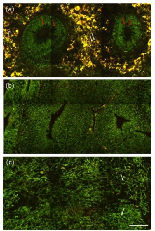
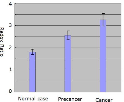
Real-time histology or virtual biopsy for the diagnosis of colonic cancer is of great medical significance. In this work, we show that label-free multiphoton imaging is feasible and effective in monitoring colonic cancer progression by providing cellular and subcellular details in fresh, unfixed, unstained colonic specimens. Our results also demonstrate the capability of using tissue quantitative analysis of the redox ratio for quantifying colonic cancer progression. These results suggest that multiphoton microscopy has potential to become an in situ histological tool, which is free from the labeling requirement of conventional methods, for the early diagnosis and detection of malignant lesions in the colon.

摘要

用于结肠癌诊断的实时组织学或虚拟活检具有重大医学意义。在这项工作中,我们表明无标记多光子成像在监测结肠癌进展方面是可行且有效的,它能在新鲜、未固定、未染色的结肠标本中提供细胞和亚细胞细节。我们的结果还证明了利用氧化还原比率的组织定量分析来量化结肠癌进展的能力。这些结果表明,多光子显微镜有潜力成为一种原位组织学工具,它无需传统方法的标记要求,可用于结肠癌恶性病变的早期诊断和检测。

https://cdn.ncbi.nlm.nih.gov/pmc/blobs/d7ce/3047366/737888bdf9d5/boe-2-3-615-g002.jpg
https://cdn.ncbi.nlm.nih.gov/pmc/blobs/d7ce/3047366/a69f0536d38f/boe-2-3-615-g001.jpg
https://cdn.ncbi.nlm.nih.gov/pmc/blobs/d7ce/3047366/737888bdf9d5/boe-2-3-615-g002.jpg
https://cdn.ncbi.nlm.nih.gov/pmc/blobs/d7ce/3047366/a69f0536d38f/boe-2-3-615-g001.jpg
https://cdn.ncbi.nlm.nih.gov/pmc/blobs/d7ce/3047366/737888bdf9d5/boe-2-3-615-g002.jpg

相似文献

1
Label-free monitoring of colonic cancer progression using multiphoton microscopy.使用多光子显微镜对结肠癌进展进行无标记监测。
Biomed Opt Express. 2011 Feb 16;2(3):615-9. doi: 10.1364/BOE.2.000615.
2
Multiphoton imaging can be used for microscopic examination of intact human gastrointestinal mucosa ex vivo.多光子成像可用于对离体的完整人类胃肠道黏膜进行显微镜检查。
Clin Gastroenterol Hepatol. 2008 Jan;6(1):95-101. doi: 10.1016/j.cgh.2007.10.008. Epub 2007 Dec 18.
3
Label-Free Monitoring of Endometrial Cancer Progression Using Multiphoton Microscopy.使用多光子显微镜进行无标记的子宫内膜癌进展监测。
Ann Biomed Eng. 2024 Nov;52(11):3113-3124. doi: 10.1007/s10439-024-03574-1. Epub 2024 Jul 3.
4
Spectrum- and time-resolved endogenous multiphoton signals reveal quantitative differentiation of premalignant and malignant gastric mucosa.光谱和时间分辨的内源性多光子信号揭示了癌前和恶性胃黏膜的定量差异。
Biomed Opt Express. 2018 Jan 8;9(2):453-471. doi: 10.1364/BOE.9.000453. eCollection 2018 Feb 1.
5
Rapid, label-free detection of intracranial germinoma using multiphoton microscopy.使用多光子显微镜对颅内生殖细胞瘤进行快速、无标记检测。
Neurophotonics. 2019 Jul;6(3):035014. doi: 10.1117/1.NPh.6.3.035014. Epub 2019 Sep 27.
6
Multiphoton microscopy: a potential intraoperative tool for the detection of carcinoma in situ in human bladder.多光子显微镜:一种用于检测人膀胱原位癌的潜在术中工具。
Arch Pathol Lab Med. 2015 Jun;139(6):796-804. doi: 10.5858/arpa.2014-0076-OA.
7
Visualization of basement membranes in normal breast and breast cancer tissues using multiphoton microscopy.利用多光子显微镜观察正常乳腺组织和乳腺癌组织中的基底膜。
Oncol Lett. 2016 Jun;11(6):3785-3789. doi: 10.3892/ol.2016.4472. Epub 2016 Apr 19.
8
Real-time histology in liver disease using multiphoton microscopy with fluorescence lifetime imaging.利用多光子显微镜和荧光寿命成像技术进行肝脏疾病的实时组织学研究。
Biomed Opt Express. 2015 Feb 10;6(3):780-92. doi: 10.1364/BOE.6.000780. eCollection 2015 Mar 1.
9
A pilot study of using multiphoton microscopy to diagnose gastric cancer.应用多光子显微镜诊断胃癌的初步研究。
Surg Endosc. 2011 May;25(5):1425-30. doi: 10.1007/s00464-010-1409-z. Epub 2010 Nov 3.
10
Multiphoton gradient index endoscopy for evaluation of diseased human prostatic tissue ex vivo.用于体外评估病变人类前列腺组织的多光子梯度折射率内窥镜检查
J Biomed Opt. 2014 Nov;19(11):116011. doi: 10.1117/1.JBO.19.11.116011.

引用本文的文献

1
Multiphoton microscopy at a microwatt level via gain-managed nonlinear amplification and pulse-picking.通过增益管理非线性放大和脉冲选择实现微瓦级多光子显微镜技术。
Biomed Opt Express. 2025 Mar 27;16(4):1692-1706. doi: 10.1364/BOE.557132. eCollection 2025 Apr 1.
2
Highly-selective optical filter for NADH fluorescence detection in multiphoton microscopy.用于多光子显微镜中烟酰胺腺嘌呤二核苷酸(NADH)荧光检测的高选择性光学滤波器。
Biomed Opt Express. 2024 Apr 24;15(5):3317-3328. doi: 10.1364/BOE.506777. eCollection 2024 May 1.
3
Automatic and label-free detection of meningioma in dura mater using the combination of multiphoton microscopy and image analysis.

本文引用的文献

1
Cancer-cell microsurgery using nonlinear optical endomicroscopy.使用非线性光学内窥显微镜进行癌细胞微创手术。
J Biomed Opt. 2010 Sep-Oct;15(5):050502. doi: 10.1117/1.3502566.
2
Time- and Spectral-resolved two-photon imaging of healthy bladder mucosa and carcinoma in situ.健康膀胱黏膜和原位癌的时间分辨与光谱分辨双光子成像
Opt Express. 2010 Feb 15;18(4):3840-9. doi: 10.1364/OE.18.003840.
3
Advances of endomicroscopy for gastrointestinal physiology and diseases.内窥式显微镜在胃肠道生理学和疾病中的应用进展。
利用多光子显微镜和图像分析相结合的方法对硬脑膜中的脑膜瘤进行自动无标记检测。
Neurophotonics. 2023 Jul;10(3):035006. doi: 10.1117/1.NPh.10.3.035006. Epub 2023 Jul 10.
4
Types of spectroscopy and microscopy techniques for cancer diagnosis: a review.用于癌症诊断的光谱学和显微镜技术类型:综述。
Lasers Med Sci. 2022 Oct;37(8):3067-3084. doi: 10.1007/s10103-022-03610-3. Epub 2022 Jul 14.
5
Spatial light interference microscopy: principle and applications to biomedicine.空间光干涉显微镜:原理及其在生物医学中的应用
Adv Opt Photonics. 2021 Jun 30;13(2):353-425. doi: 10.1364/AOP.417837. Epub 2021 May 5.
6
Diagnosing Hirschsprung disease by detecting intestinal ganglion cells using label-free hyperspectral microscopy.利用无标记高光谱显微镜检测肠神经节细胞诊断先天性巨结肠症。
Sci Rep. 2021 Jan 14;11(1):1398. doi: 10.1038/s41598-021-80981-7.
7
Margin diagnosis for endoscopic submucosal dissection of early gastric cancer using multiphoton microscopy.应用多光子显微镜对早期胃癌内镜黏膜下剥离术的边缘诊断。
Surg Endosc. 2020 Jan;34(1):408-416. doi: 10.1007/s00464-019-06783-1. Epub 2019 Apr 10.
8
Epithelial Ovarian Cancer Diagnosis of Second-Harmonic Generation Images: A Semiautomatic Collagen Fibers Quantification Protocol.上皮性卵巢癌二次谐波生成图像的诊断:一种半自动胶原纤维定量方案
Cancer Inform. 2017 Feb 3;16:1176935117690162. doi: 10.1177/1176935117690162. eCollection 2017.
9
Biophotonic endoscopy: a review of clinical research techniques for optical imaging and sensing of early gastrointestinal cancer.生物光子内镜检查:早期胃肠道癌光学成像与传感临床研究技术综述
Endosc Int Open. 2015 Oct;3(5):E380-92. doi: 10.1055/s-0034-1392513. Epub 2015 Sep 8.
10
In vivo nonlinear spectral imaging as a tool to monitor early spectroscopic and metabolic changes in a murine cutaneous squamous cell carcinoma model.体内非线性光谱成像作为监测小鼠皮肤鳞状细胞癌模型早期光谱和代谢变化的工具。
Biomed Opt Express. 2014 Nov 13;5(12):4281-99. doi: 10.1364/BOE.5.004281. eCollection 2014 Dec 1.
Am J Physiol Gastrointest Liver Physiol. 2010 Jun;298(6):G797-806. doi: 10.1152/ajpgi.00027.2010. Epub 2010 Feb 25.
4
Multimode nonlinear optical imaging of the dermis in ex vivo human skin based on the combination of multichannel mode and Lambda mode.基于多通道模式和Lambda模式相结合的离体人皮肤真皮多模非线性光学成像
Opt Express. 2006 Aug 21;14(17):7810-20. doi: 10.1364/oe.14.007810.
5
Cancer statistics, 2009.2009年癌症统计数据。
CA Cancer J Clin. 2009 Jul-Aug;59(4):225-49. doi: 10.3322/caac.20006. Epub 2009 May 27.
6
Extracting diagnostic stromal organization features based on intrinsic two-photon excited fluorescence and second-harmonic generation signals.基于双光子激发荧光和二次谐波信号提取诊断性基质组织特征。
J Biomed Opt. 2009 Mar-Apr;14(2):020503. doi: 10.1117/1.3088029.
7
Multiphoton imaging can be used for microscopic examination of intact human gastrointestinal mucosa ex vivo.多光子成像可用于对离体的完整人类胃肠道黏膜进行显微镜检查。
Clin Gastroenterol Hepatol. 2008 Jan;6(1):95-101. doi: 10.1016/j.cgh.2007.10.008. Epub 2007 Dec 18.
8
In vivo multiphoton microscopy of NADH and FAD redox states, fluorescence lifetimes, and cellular morphology in precancerous epithelia.癌前上皮中烟酰胺腺嘌呤二核苷酸(NADH)和黄素腺嘌呤二核苷酸(FAD)氧化还原状态、荧光寿命及细胞形态的体内多光子显微镜检查
Proc Natl Acad Sci U S A. 2007 Dec 4;104(49):19494-9. doi: 10.1073/pnas.0708425104. Epub 2007 Nov 27.
9
Clinical two-photon microendoscopy.临床双光子显微内镜检查
Microsc Res Tech. 2007 May;70(5):398-402. doi: 10.1002/jemt.20445.
10
Multiphoton microscopy of endogenous fluorescence differentiates normal, precancerous, and cancerous squamous epithelial tissues.内源性荧光的多光子显微镜可区分正常、癌前和癌性鳞状上皮组织。
Cancer Res. 2005 Feb 15;65(4):1180-6. doi: 10.1158/0008-5472.CAN-04-3031.