Neuchrist C, Grasl M, Scheiner O, Lassmann H, Ehrenberger K, Kraft D
Institute of General and Experimental Pathology, Vienna, Austria.
Br J Cancer. 1990 Nov;62(5):748-53. doi: 10.1038/bjc.1990.371.
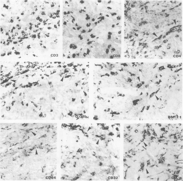
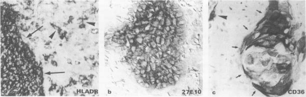
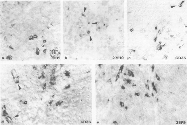
Biopsies from 26 patients with advanced stage squamous cell carcinoma of the head and neck were investigated to determine the intensity of the inflammatory cellular infiltrate and the expression of leucocyte antigens. Mononuclear cell infiltration varied considerably between the individual patients and also within the tumour. Tumour-infiltrating cells consisted mainly of T lymphocytes and monocytes (Mo)/macrophages (M phi). Staining procedure with monoclonal antibodies (moabs) against Mo/M phi revealed different clusters of antigen expression: (1) moabs 27E10 and a-CD35 detected a subgroup of Mo/M phi with particular staining of perivasal Mo; (2) moab a-CD1 stained preferentially cells in tumour cell clusters; (3) moabs that reacted with cells of either typical M phi or dendritic morphology throughout the tumour-tissue: a-Fc gamma receptor I-III, a-class II antigens, a-CD4, Rm3/1, a-CD36 and 25F9. Thus, the majority of tumour-infiltrating mononuclear phagocytes were found to possess a rather mature phenotype. The number of Mo/M phi with mature phenotype within the tumours correlated with T lymphocyte infiltration in the tissue.
对26例晚期头颈部鳞状细胞癌患者的活检组织进行研究,以确定炎性细胞浸润的强度和白细胞抗原的表达。单核细胞浸润在个体患者之间以及肿瘤内部均有很大差异。肿瘤浸润细胞主要由T淋巴细胞和单核细胞(Mo)/巨噬细胞(M phi)组成。用抗Mo/M phi的单克隆抗体(moabs)进行染色,发现了不同的抗原表达簇:(1)moabs 27E10和α-CD35检测到Mo/M phi的一个亚群,其血管周围的Mo有特殊染色;(2)α-CD1单克隆抗体优先染色肿瘤细胞簇中的细胞;(3)在整个肿瘤组织中与典型M phi或树突状形态细胞反应的单克隆抗体:α-Fcγ受体I-III、α-Ⅱ类抗原、α-CD4、Rm3/1、α-CD36和25F9。因此,发现大多数肿瘤浸润性单核吞噬细胞具有相当成熟的表型。肿瘤内具有成熟表型的Mo/M phi数量与组织中的T淋巴细胞浸润相关。