Ferrer Consuelo, Alió Jorge L
J Ophthalmic Inflamm Infect. 2011 Feb 23;1(1):15-22. doi: 10.1007/s12348-011-0019-9.
The aims of this study were to assess the utility of polymerase chain reaction (PCR) in diagnosing fungal keratitis in the last decade in our center and to review the molecular diagnosis of mycotic keratitis.
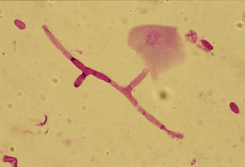
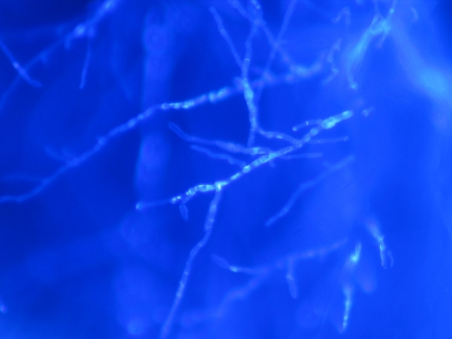
A retrospective nonrandomized investigation was undertaken at Vissum Corporación Instituto Oftalmologico de Alicante to evaluate 27 corneal samples of 20 patients with proven fungal keratitis from January 2000 to December 2009. Corneal samples (21 corneal scrapings, 5 biopsies, and 1 cornea) were evaluated by Gram stain or calcofluor stain, culture, and PCR. The detection and molecular identification were carried out by DNA amplification and sequencing of the internal transcribed spacer and 5.8S rRNA region from the corneal samples.
PCR detected all the samples that were positive by conventional methods. Four samples were positive by PCR and showed negative results by culture and stain. Combination of microscopy and culture gave positive results in 21 of the 27 samples of patients with mycotic keratitis. Stains showed a 66.7% of positive results, culture showed 59.3%, and PCR showed 92.6%. The time taken for PCR assay was 4 to 8 h whereas positive fungal cultures took 1 to 35 days. Identification at species level by molecular methods was possible in all cases except one. Identification at species level by conventional methods only was possible in eight cases.
PCR not only proved to be an effective rapid method for the diagnosis of fungal keratitis but was also more sensitive than stain and culture methods. Fungal PCR must be added as the screening diagnosis test when an early mycotic keratitis is suspected. Molecular identification is the gold standard technique for the identification of corneal fungal pathogens.
本研究旨在评估聚合酶链反应(PCR)在过去十年中于我们中心诊断真菌性角膜炎的效用,并回顾真菌性角膜炎的分子诊断。
在阿利坎特眼科研究所维苏姆公司进行了一项回顾性非随机调查,以评估2000年1月至2009年12月期间20例经证实患有真菌性角膜炎患者的27份角膜样本。角膜样本(21份角膜刮片、5份活检样本和1个角膜)通过革兰氏染色或荧光增白剂染色、培养和PCR进行评估。通过对角膜样本的内部转录间隔区和5.8S rRNA区域进行DNA扩增和测序来进行检测和分子鉴定。
PCR检测出所有通过传统方法呈阳性的样本。4份样本经PCR检测呈阳性,但培养和染色结果为阴性。显微镜检查和培养相结合在27份真菌性角膜炎患者样本中的21份中得出阳性结果。染色显示阳性结果的比例为66.7%,培养显示为59.3%,而PCR显示为92.6%。PCR检测所需时间为4至8小时,而真菌培养阳性则需要1至35天。除1例病例外,所有病例均有可能通过分子方法在种属水平进行鉴定。仅通过传统方法在种属水平进行鉴定的有8例。
PCR不仅被证明是诊断真菌性角膜炎的一种有效快速方法,而且比染色和培养方法更敏感。当怀疑早期真菌性角膜炎时,必须将真菌PCR作为筛查诊断试验。分子鉴定是鉴定角膜真菌病原体的金标准技术。