Unit of Infection Models, German Primate Center, Leibniz Institute for Primate Research, Kellnerweg 4, 37077 Goettingen, Germany.
Retrovirology. 2011 Apr 11;8:24. doi: 10.1186/1742-4690-8-24.
Since there is still no protective HIV vaccine available, better insights into immune mechanism of persons effectively controlling HIV replication in the absence of any therapy should contribute to improve further vaccine designs. However, little is known about the mucosal immune response of this small unique group of patients. Using the SIV-macaque-model for AIDS, we had the rare opportunity to analyze 14 SIV-infected rhesus macaques durably controlling viral replication (controllers). We investigated the virological and immunological profile of blood and three different mucosal tissues and compared their data to those of uninfected and animals progressing to AIDS-like disease (progressors).
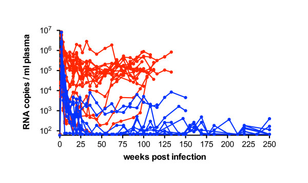
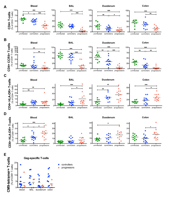
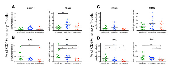
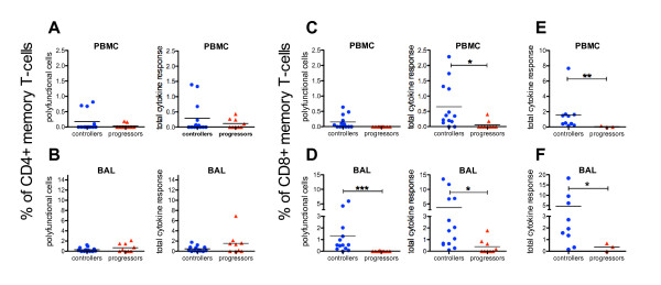
Lymphocytes from blood, bronchoalveolar lavage (BAL), and duodenal and colonic biopsies were phenotypically characterized by polychromatic flow cytometry. In controllers, we observed higher levels of CD4+, CD4+CCR5+ and Gag-specific CD8+ T-cells as well as lower immune activation in blood and all mucosal sites compared to progressors. However, we could also demonstrate that immunological changes are distinct between these three mucosal sites.Intracellular cytokine staining demonstrated a significantly higher systemic and mucosal CD8+ Gag-specific cellular immune response in controllers than in progressors. Most remarkable was the polyfunctional cytokine profile of CD8+ lymphocytes in BAL of controllers, which significantly dominated over their blood response. The overall suppression of viral replication in the controllers was confirmed by almost no detectable viral RNA in blood and all mucosal tissues investigated.
A strong and complex virus-specific CD8+ T-cell response in blood and especially in mucosal tissue of SIV-infected macaques was associated with low immune activation and an efficient suppression of viral replication. This likely afforded a repopulation of CD4+ T-cells in different mucosal compartments to almost normal levels. We conclude, that a robust SIV-specific mucosal immune response seems to be essential for establishing and maintaining the controller status and consequently for long-term survival.
由于目前尚无有效的 HIV 保护疫苗,因此深入了解在没有任何治疗的情况下有效控制 HIV 复制的个体的免疫机制,有助于进一步改进疫苗设计。然而,人们对这一小部分独特患者的黏膜免疫反应知之甚少。利用 SIV-猕猴艾滋病模型,我们有机会分析了 14 只长期控制病毒复制的 SIV 感染猕猴(控制者)。我们研究了血液和三种不同黏膜组织的病毒学和免疫学特征,并将其数据与未感染和进展为艾滋病样疾病的动物(进展者)进行了比较。
通过多色流式细胞术对血液、支气管肺泡灌洗液(BAL)、十二指肠和结肠活检的淋巴细胞进行了表型特征分析。与进展者相比,控制者血液和所有黏膜部位的 CD4+、CD4+CCR5+和 Gag 特异性 CD8+T 细胞水平更高,免疫激活水平更低。然而,我们也证明了这些三个黏膜部位的免疫变化是不同的。细胞内细胞因子染色显示,与进展者相比,控制者的系统和黏膜 Gag 特异性 CD8+细胞免疫反应明显更高。最显著的是控制者 BAL 中 CD8+淋巴细胞的多效细胞因子谱,明显超过其血液反应。控制者体内几乎检测不到血液和所有研究黏膜组织中的病毒 RNA,证实了病毒复制的总体抑制。
在 SIV 感染猕猴的血液和特别是黏膜组织中,强烈而复杂的病毒特异性 CD8+T 细胞反应与低免疫激活和高效抑制病毒复制有关。这可能使不同黏膜部位的 CD4+T 细胞重新得到补充,接近正常水平。我们得出结论,强大的 SIV 特异性黏膜免疫反应似乎是建立和维持控制者状态以及长期生存的必要条件。