• 文献检索
  • 文档翻译
  • 深度研究
  • 学术资讯
  • Suppr Zotero 插件Zotero 插件
  • 邀请有礼
  • 套餐&价格
  • 历史记录
应用&插件
Suppr Zotero 插件Zotero 插件浏览器插件Mac 客户端Windows 客户端微信小程序
定价
高级版会员购买积分包购买API积分包
服务
文献检索文档翻译深度研究API 文档MCP 服务
关于我们
关于 Suppr公司介绍联系我们用户协议隐私条款
关注我们

Suppr 超能文献

核心技术专利:CN118964589B侵权必究
粤ICP备2023148730 号-1Suppr @ 2026

文献检索

告别复杂PubMed语法,用中文像聊天一样搜索,搜遍4000万医学文献。AI智能推荐,让科研检索更轻松。

立即免费搜索

文件翻译

保留排版,准确专业,支持PDF/Word/PPT等文件格式,支持 12+语言互译。

免费翻译文档

深度研究

AI帮你快速写综述,25分钟生成高质量综述,智能提取关键信息,辅助科研写作。

立即免费体验

使用具有自适应光学的超高分辨率光学相干断层扫描在三维空间和时间上对视锥光感受器进行成像。

Imaging cone photoreceptors in three dimensions and in time using ultrahigh resolution optical coherence tomography with adaptive optics.

作者信息

Kocaoglu Omer P, Lee Sangyeol, Jonnal Ravi S, Wang Qiang, Herde Ashley E, Derby Jack C, Gao Weihua, Miller Donald T

机构信息

School of Optometry, Indiana University, 800 East Atwater Avenue, Bloomington, IN 47405, USA.

出版信息

Biomed Opt Express. 2011 Mar 1;2(4):748-63. doi: 10.1364/BOE.2.000748.

DOI:10.1364/BOE.2.000748
PMID:21483600
原文链接:https://pmc.ncbi.nlm.nih.gov/articles/PMC3072118/
Abstract

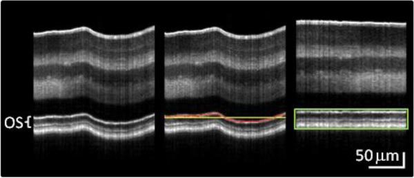
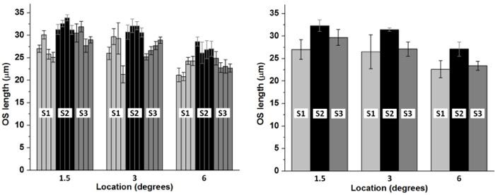
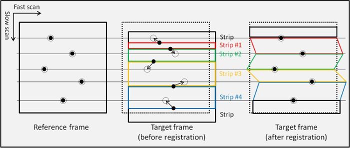
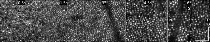
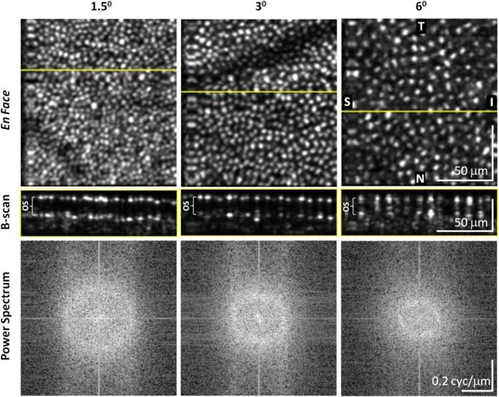
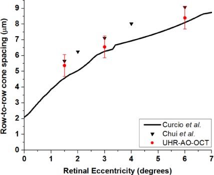
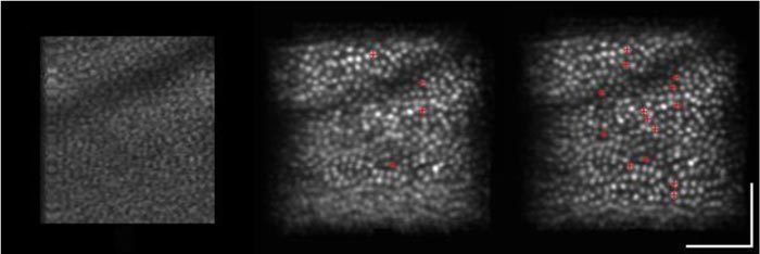
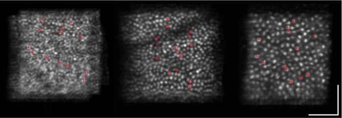
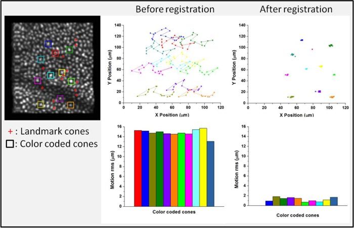
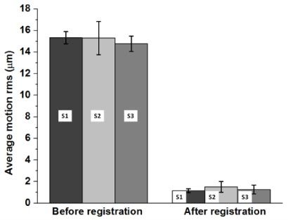
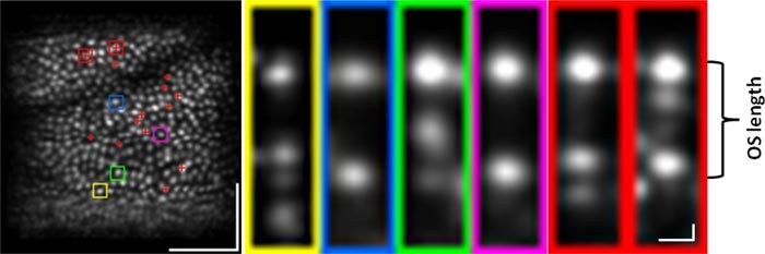
Cone photoreceptors in the living human eye have recently been imaged with micron-scale resolution in all three spatial dimensions using adaptive optics optical coherence tomography. While these advances have allowed non-invasive study of the three-dimensional structure of living human cones, studies of their function and physiology are still hampered by the difficulties to monitor the same cells over time. The purpose of this study is to demonstrate the feasibility of cone monitoring using ultrahigh-resolution adaptive optics optical coherence tomography. Critical to this is incorporation of a high speed CMOS camera (125 KHz) and a novel feature-based, image registration/dewarping algorithm for reducing the deleterious effects of eye motion on volume images. Volume movies were acquired on three healthy subjects at retinal eccentricities from 0.5° to 6°. Image registration/dewarping reduced motion artifacts in the movies from 15 μm to 1.3 μm root mean square, the latter sufficient for identifying and tracking cones. Cone row-to-row spacing and outer segment lengths were consistent with that reported in the literature. Cone length analysis demonstrates that UHR-AO-OCT is sufficiently sensitive to measure real length differences between cones in the same 0.5° retinal patch, and requires no more than five measurements of OS length to achieve 95% confidence. We know of no other imaging modality that can monitor foveal or parafoveal cones over time with comparable resolution in all three dimensions.

摘要

最近,利用自适应光学光学相干断层扫描技术,已在三个空间维度上以微米级分辨率对活人眼中的视锥光感受器进行了成像。虽然这些进展使得对活人视锥细胞的三维结构进行非侵入性研究成为可能,但对视锥细胞功能和生理学的研究仍因难以长时间监测同一细胞而受到阻碍。本研究的目的是证明使用超高分辨率自适应光学光学相干断层扫描技术监测视锥细胞的可行性。对此至关重要的是采用高速CMOS相机(125千赫兹)和一种基于特征的新型图像配准/去扭曲算法,以减少眼球运动对体图像的有害影响。在三名健康受试者的视网膜偏心度为0.5°至6°的区域采集了体视电影。图像配准/去扭曲将电影中的运动伪影从均方根15微米减少到1.3微米,后者足以识别和跟踪视锥细胞。视锥细胞的行间距和外段长度与文献报道一致。视锥细胞长度分析表明,超高分辨率自适应光学光学相干断层扫描技术足够灵敏,能够测量同一0.5°视网膜区域内视锥细胞之间的实际长度差异,并且测量视锥细胞外段长度不超过五次就能达到95%的置信度。我们所知的其他成像方式,都无法在所有三个维度上以可比的分辨率长时间监测中央凹或中央凹旁的视锥细胞。

https://cdn.ncbi.nlm.nih.gov/pmc/blobs/d31d/3072118/577a53848194/boe-2-4-748-g011.jpg
https://cdn.ncbi.nlm.nih.gov/pmc/blobs/d31d/3072118/cf282e3176d6/boe-2-4-748-g001.jpg
https://cdn.ncbi.nlm.nih.gov/pmc/blobs/d31d/3072118/733a498f8bf0/boe-2-4-748-g002.jpg
https://cdn.ncbi.nlm.nih.gov/pmc/blobs/d31d/3072118/dae217854575/boe-2-4-748-g003.jpg
https://cdn.ncbi.nlm.nih.gov/pmc/blobs/d31d/3072118/04a3d1383522/boe-2-4-748-g004.jpg
https://cdn.ncbi.nlm.nih.gov/pmc/blobs/d31d/3072118/8e24d45e5e76/boe-2-4-748-g005.jpg
https://cdn.ncbi.nlm.nih.gov/pmc/blobs/d31d/3072118/c0b03f5f4ada/boe-2-4-748-g006.jpg
https://cdn.ncbi.nlm.nih.gov/pmc/blobs/d31d/3072118/a26f35679faa/boe-2-4-748-g007.jpg
https://cdn.ncbi.nlm.nih.gov/pmc/blobs/d31d/3072118/3199f5cf1844/boe-2-4-748-g008.jpg
https://cdn.ncbi.nlm.nih.gov/pmc/blobs/d31d/3072118/f638b583d2ab/boe-2-4-748-g009.jpg
https://cdn.ncbi.nlm.nih.gov/pmc/blobs/d31d/3072118/bd927534aa9e/boe-2-4-748-g010.jpg
https://cdn.ncbi.nlm.nih.gov/pmc/blobs/d31d/3072118/577a53848194/boe-2-4-748-g011.jpg

相似文献

1
Imaging cone photoreceptors in three dimensions and in time using ultrahigh resolution optical coherence tomography with adaptive optics.使用具有自适应光学的超高分辨率光学相干断层扫描在三维空间和时间上对视锥光感受器进行成像。
Biomed Opt Express. 2011 Mar 1;2(4):748-63. doi: 10.1364/BOE.2.000748.
2
Morphologic and Functional Assessment of Photoreceptors After Macula-Off Retinal Detachment With Adaptive-Optics OCT and Microperimetry.黄斑裂孔性视网膜脱离后应用自适应光学 OCT 和微视野计对光感受器的形态和功能评估。
Am J Ophthalmol. 2020 Jun;214:72-85. doi: 10.1016/j.ajo.2019.12.015. Epub 2019 Dec 25.
3
Kilohertz retinal FF-SS-OCT and flood imaging with hardware-based adaptive optics.千赫兹视网膜频域分段扫描光学相干断层扫描与基于硬件自适应光学的泛光成像
Biomed Opt Express. 2020 Sep 30;11(10):5995-6011. doi: 10.1364/BOE.403509. eCollection 2020 Oct 1.
4
Three-dimensional composition of the photoreceptor cone layers in healthy eyes using adaptive-optics optical coherence tomography (AO-OCT).利用自适应光学光相干断层扫描(AO-OCT)技术对健康眼的光感受器锥体细胞层进行三维成像。
PLoS One. 2021 Jan 7;16(1):e0245293. doi: 10.1371/journal.pone.0245293. eCollection 2021.
5
Evaluation of Foveal Cone and Müller Cells in Epiretinal Membrane using Adaptive Optics OCT.使用自适应光学光学相干断层扫描技术评估视网膜前膜中的中央凹视锥细胞和米勒细胞。
Ophthalmol Sci. 2023 Jul 4;4(1):100362. doi: 10.1016/j.xops.2023.100362. eCollection 2024 Jan-Feb.
6
High-speed volumetric imaging of cone photoreceptors with adaptive optics spectral-domain optical coherence tomography.利用自适应光学光谱域光学相干断层扫描技术对视锥光感受器进行高速容积成像。
Opt Express. 2006 May 15;14(10):4380-94. doi: 10.1364/OE.14.004380.
7
The cellular origins of the outer retinal bands in optical coherence tomography images.光学相干断层扫描图像中外侧视网膜带的细胞起源
Invest Ophthalmol Vis Sci. 2014 Oct 16;55(12):7904-18. doi: 10.1167/iovs.14-14907.
8
Ultrahigh speed spectral / Fourier domain OCT ophthalmic imaging at 70,000 to 312,500 axial scans per second.每秒70,000至312,500次轴向扫描的超高速光谱/傅里叶域光学相干断层扫描眼科成像。
Opt Express. 2008 Sep 15;16(19):15149-69. doi: 10.1364/oe.16.015149.
9
Three-dimensional assessment of para- and perifoveal photoreceptor densities and the impact of meridians and age in healthy eyes with adaptive-optics optical coherence tomography (AO-OCT).应用自适应光学 OCT 对健康眼的旁中心和中心凹周围视锥细胞密度的三维评估及其与子午线和年龄的关系。
Opt Express. 2020 Nov 23;28(24):36723-36739. doi: 10.1364/OE.409076.
10
Automated identification of cone photoreceptors in adaptive optics optical coherence tomography images using transfer learning.利用迁移学习在自适应光学光学相干断层扫描图像中自动识别视锥光感受器。
Biomed Opt Express. 2018 Oct 10;9(11):5353-5367. doi: 10.1364/BOE.9.005353. eCollection 2018 Nov 1.

引用本文的文献

1
Evolution of adaptive optics retinal imaging [Invited].自适应光学视网膜成像的发展[特邀报告]
Biomed Opt Express. 2023 Feb 28;14(3):1307-1338. doi: 10.1364/BOE.485371. eCollection 2023 Mar 1.
2
Deep learning-enabled volumetric cone photoreceptor segmentation in adaptive optics optical coherence tomography images of normal and diseased eyes.在正常和患病眼睛的自适应光学光学相干断层扫描图像中,基于深度学习的体积视锥光感受器分割
Biomed Opt Express. 2023 Jan 23;14(2):815-833. doi: 10.1364/BOE.478693. eCollection 2023 Feb 1.
3
The Development and Clinical Application of Innovative Optical Ophthalmic Imaging Techniques.

本文引用的文献

1
Design of an integrated hardware interface for AOSLO image capture and cone-targeted stimulus delivery.用于自适应光学扫描激光眼科显微镜(AOSLO)图像采集和视锥靶向刺激传递的集成硬件接口设计
Opt Express. 2010 Aug 16;18(17):17841-58. doi: 10.1364/OE.18.017841.
2
In vivo investigation of human cone photoreceptors with SLO/OCT in combination with 3D motion correction on a cellular level.利用SLO/OCT结合细胞水平的三维运动校正对人类视锥光感受器进行体内研究。
Opt Express. 2010 Jun 21;18(13):13935-44. doi: 10.1364/OE.18.013935.
3
Imaging outer segment renewal in living human cone photoreceptors.
创新光学眼科成像技术的发展与临床应用
Front Med (Lausanne). 2022 Jun 30;9:891369. doi: 10.3389/fmed.2022.891369. eCollection 2022.
4
Multi-modal and multi-scale clinical retinal imaging system with pupil and retinal tracking.具有瞳孔和视网膜跟踪的多模态和多尺度临床视网膜成像系统。
Sci Rep. 2022 Jun 10;12(1):9577. doi: 10.1038/s41598-022-13631-1.
5
Advances in multimodal imaging in ophthalmology.眼科多模态成像的进展。
Ther Adv Ophthalmol. 2021 Mar 19;13:25158414211002400. doi: 10.1177/25158414211002400. eCollection 2021 Jan-Dec.
6
Toward a clinical optoretinogram: a review of noninvasive, optical tests of retinal neural function.迈向临床视网膜电图:视网膜神经功能的非侵入性光学检测综述
Ann Transl Med. 2021 Aug;9(15):1270. doi: 10.21037/atm-20-6440.
7
Bardet-Biedl syndrome-7 () shows treatment potential and a cone-rod dystrophy phenotype that recapitulates the non-human primate model.Bardet-Biedl 综合征-7()显示出治疗潜力和圆锥-杆状细胞营养不良表型,可再现非人类灵长类动物模型。
Ophthalmic Genet. 2021 Jun;42(3):252-265. doi: 10.1080/13816810.2021.1888132. Epub 2021 Mar 17.
8
Three-dimensional composition of the photoreceptor cone layers in healthy eyes using adaptive-optics optical coherence tomography (AO-OCT).利用自适应光学光相干断层扫描(AO-OCT)技术对健康眼的光感受器锥体细胞层进行三维成像。
PLoS One. 2021 Jan 7;16(1):e0245293. doi: 10.1371/journal.pone.0245293. eCollection 2021.
9
Multi-reference global registration of individual A-lines in adaptive optics optical coherence tomography retinal images.多参考个体 A 线自适应光学光相干断层扫描视网膜图像的全局配准。
J Biomed Opt. 2021 Jan;26(1). doi: 10.1117/1.JBO.26.1.016001.
10
High-Speed, Ultrahigh-Resolution Spectral-Domain OCT with Extended Imaging Range Using Reference Arm Length Matching.高速、超高分辨率谱域 OCT 采用参考臂长匹配技术扩展成像范围。
Transl Vis Sci Technol. 2020 Jun 8;9(7):12. doi: 10.1167/tvst.9.7.12. eCollection 2020 Jun.
活体人类视锥光感受器中外段更新的成像
Opt Express. 2010 Mar 1;18(5):5257-70. doi: 10.1364/OE.18.005257.
4
Arrested development: high-resolution imaging of foveal morphology in albinism.发育停滞:白化病中黄斑形态的高分辨率成像
Vision Res. 2010 Apr 7;50(8):810-7. doi: 10.1016/j.visres.2010.02.003. Epub 2010 Feb 10.
5
Retinal imaging with polarization-sensitive optical coherence tomography and adaptive optics.使用偏振敏感光学相干断层扫描和自适应光学技术的视网膜成像。
Opt Express. 2009 Nov 23;17(24):21634-51. doi: 10.1364/OE.17.021634.
6
Adaptive optics optical coherence tomography at 120,000 depth scans/s for non-invasive cellular phenotyping of the living human retina.用于活体人类视网膜非侵入性细胞表型分析的每秒120,000次深度扫描的自适应光学光学相干断层扫描技术
Opt Express. 2009 Oct 26;17(22):19382-400. doi: 10.1364/OE.17.019382.
7
Simultaneous SLO/OCT imaging of the human retina with axial eye motion correction.具有轴向眼球运动校正功能的人视网膜同步扫描激光眼底镜/光学相干断层扫描成像
Opt Express. 2007 Dec 10;15(25):16922-32. doi: 10.1364/oe.15.016922.
8
Chromatic aberration correction of the human eye for retinal imaging in the near infrared.人眼在近红外视网膜成像中的色差校正。
Opt Express. 2006 Jun 26;14(13):6213-25. doi: 10.1364/oe.14.006213.
9
Adaptive optics flood-illumination camera for high speed retinal imaging.用于高速视网膜成像的自适应光学泛光照明相机。
Opt Express. 2006 May 15;14(10):4552-69. doi: 10.1364/oe.14.004552.
10
Adaptive optics scanning laser ophthalmoscope for stabilized retinal imaging.用于稳定视网膜成像的自适应光学扫描激光检眼镜。
Opt Express. 2006 Apr 17;14(8):3354-67. doi: 10.1364/oe.14.003354.