Rheumatology and Clinical Immunology, University Medical Center Utrecht, Utrecht, The Netherlands.
Ann Rheum Dis. 2011 Aug;70(8):1441-6. doi: 10.1136/ard.2010.142364. Epub 2011 May 12.
Modification of joint tissue damage is challenging in late-stage osteoarthritis (OA). Few options are available for treating end-stage knee OA other than joint replacement.
To examine whether joint distraction can effectively modify knee joint tissue damage and has the potential to delay prosthesis surgery.
20 patients (<60 years) with tibiofemoral OA were treated surgically using joint distraction. Distraction (~5 mm) was applied for 2 months using an external fixation frame. Tissue structure modification at 1 year of follow-up was evaluated radiographically (joint space width (JSW)), by MRI (segmentation of cartilage morphology) and by biochemical markers of collagen type II turnover, with operators blinded to time points. Clinical improvement was evaluated by Western Ontario and McMaster Universities Osteoarthritis Index (WOMAC) and Visual Analogue Scale (VAS) pain score.
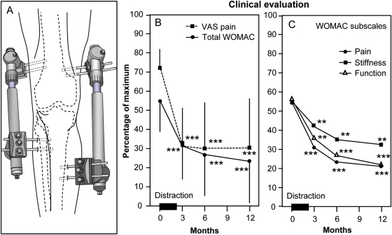
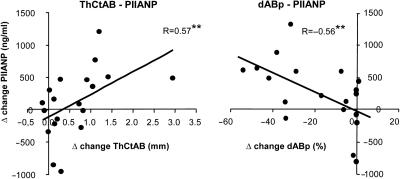
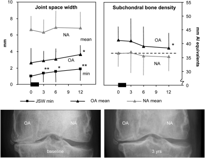
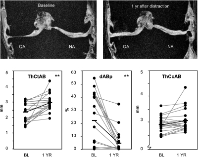
Radiography demonstrated an increase in mean and minimum JSW (2.7 to 3.6 mm and 1.0 to 1.9 mm; p<0.05 and <0.01). MRI revealed an increase in cartilage thickness (2.4 to 3.0 mm; p<0.001) and a decrease of denuded bone areas (22% to 5%; p<0.001). Collagen type II levels showed a trend towards increased synthesis (+103%; p<0.06) and decreased breakdown (-11%; p<0.08). The WOMAC index increased from 45 to 77 points, and VAS pain decreased from 73 to 31 mm (both p<0.001).
Joint distraction can induce tissue structure modification in knee OA and could result in clinical benefit. No current treatment is able to induce such changes. Larger, longer and randomised studies on joint distraction are warranted.
晚期骨关节炎(OA)的关节组织损伤难以修复。除了关节置换,治疗终末期膝骨关节炎的选择有限。
研究关节牵张术是否能有效改善膝关节组织损伤,是否有潜力延迟假体手术。
对 20 例(<60 岁)胫股 OA 患者采用关节牵张术进行手术治疗。使用外固定架进行为期 2 个月的牵张(~5mm)。在 1 年的随访时,通过影像学(关节间隙宽度(JSW))、MRI(软骨形态分割)和 II 型胶原转换的生化标志物评估组织结构的改变,操作者对时间点不知情。采用 Western Ontario 和 McMaster 大学骨关节炎指数(WOMAC)和视觉模拟评分(VAS)疼痛评分评估临床改善情况。
影像学显示平均和最小 JSW 增加(2.7 至 3.6mm 和 1.0 至 1.9mm;p<0.05 和 p<0.01)。MRI 显示软骨厚度增加(2.4 至 3.0mm;p<0.001)和裸露骨面积减少(22%至 5%;p<0.001)。II 型胶原水平显示合成增加(+103%;p<0.06)和分解减少(-11%;p<0.08)的趋势。WOMAC 指数从 45 分增加到 77 分,VAS 疼痛从 73 分降低到 31 分(均 p<0.001)。
关节牵张术可引起膝骨关节炎的组织结构改变,并可能带来临床获益。目前尚无任何治疗方法能够诱导这种改变。需要进行更大、更长时间和随机对照研究来验证关节牵张术的效果。