• 文献检索
  • 文档翻译
  • 深度研究
  • 学术资讯
  • Suppr Zotero 插件Zotero 插件
  • 邀请有礼
  • 套餐&价格
  • 历史记录
应用&插件
Suppr Zotero 插件Zotero 插件浏览器插件Mac 客户端Windows 客户端微信小程序
定价
高级版会员购买积分包购买API积分包
服务
文献检索文档翻译深度研究API 文档MCP 服务
关于我们
关于 Suppr公司介绍联系我们用户协议隐私条款
关注我们

Suppr 超能文献

核心技术专利:CN118964589B侵权必究
粤ICP备2023148730 号-1Suppr @ 2026

文献检索

告别复杂PubMed语法,用中文像聊天一样搜索,搜遍4000万医学文献。AI智能推荐,让科研检索更轻松。

立即免费搜索

文件翻译

保留排版,准确专业,支持PDF/Word/PPT等文件格式,支持 12+语言互译。

免费翻译文档

深度研究

AI帮你快速写综述,25分钟生成高质量综述,智能提取关键信息,辅助科研写作。

立即免费体验

三维超声心动图左心室定量分析:基础验证与临床应用。

Three-dimensional echocardiography for left ventricular quantification: fundamental validation and clinical applications.

机构信息

Department of Cardiology 5F 003, VU University Medical Center, De Boelelaan 1117, 1081 HV, Amsterdam, the Netherlands.

出版信息

Neth Heart J. 2011 Oct;19(10):423-31. doi: 10.1007/s12471-011-0160-y.

DOI:10.1007/s12471-011-0160-y
PMID:21584798
原文链接:https://pmc.ncbi.nlm.nih.gov/articles/PMC3189315/
Abstract

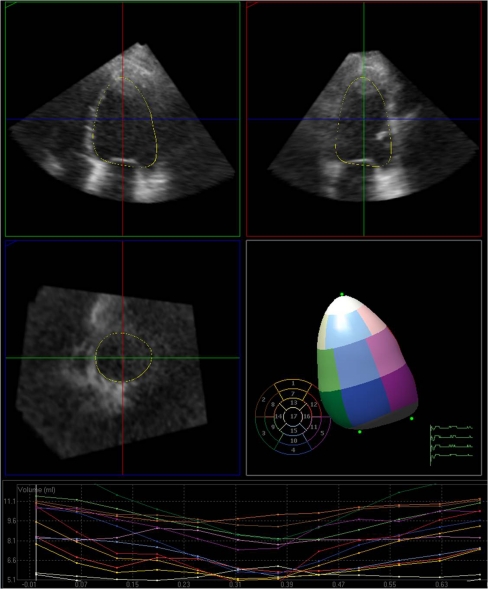
One of the earliest applications of clinical echocardiography is evaluation of left ventricular (LV) function and size. Accurate, reproducible and quantitative evaluation of LV function and size is vital for diagnosis, treatment and prediction of prognosis of heart disease. Early three-dimensional (3D) echocardiographic techniques showed better reproducibility than two-dimensional (2D) echocardiography and narrower limits of agreement for assessment of LV function and size in comparison to reference methods, mostly cardiac magnetic resonance (CMR) imaging, but acquisition methods were cumbersome and a lack of user-friendly analysis software initially precluded widespread use. Through the advent of matrix transducers enabling real-time three-dimensional echocardiography (3DE) and improvements in analysis software featuring semi-automated volumetric analysis, 3D echocardiography evolved into a simple and fast imaging modality for everyday clinical use. 3DE provides the possibility to evaluate the entire LV in three spatial dimensions during the complete cardiac cycle, offering a more accurate and complete quantitative evaluation the LV. Improved efficiency in acquisition and analysis may provide clinicians with important diagnostic information within minutes. The current article reviews the methodology and application of 3DE for quantitative evaluation of the LV, provides the scientific evidence for its current clinical use, and discusses its current limitations and potential future directions.

摘要

临床超声心动图最早的应用之一是评估左心室(LV)功能和大小。准确、可重复和定量评估 LV 功能和大小对于心脏病的诊断、治疗和预后预测至关重要。早期的三维(3D)超声心动图技术显示出比二维(2D)超声心动图更好的可重复性,并且与参考方法(主要是心脏磁共振成像(CMR)成像)相比,评估 LV 功能和大小的协议限制更窄,但采集方法繁琐,缺乏用户友好的分析软件,最初限制了其广泛应用。随着能够实现实时三维超声心动图(3DE)的矩阵换能器的出现以及具有半自动容积分析功能的分析软件的改进,3D 超声心动图已发展成为一种简单快捷的日常临床成像方式。3DE 可在整个心动周期内从三个空间维度评估整个 LV,提供更准确和完整的 LV 定量评估。在采集和分析方面效率的提高可能会在数分钟内为临床医生提供重要的诊断信息。本文综述了 3DE 定量评估 LV 的方法学和应用,提供了其当前临床应用的科学证据,并讨论了其当前的局限性和潜在的未来方向。

https://cdn.ncbi.nlm.nih.gov/pmc/blobs/4d0b/3189315/94f9b2c37f4a/12471_2011_160_Fig3_HTML.jpg
https://cdn.ncbi.nlm.nih.gov/pmc/blobs/4d0b/3189315/4300cac8db81/12471_2011_160_Fig1_HTML.jpg
https://cdn.ncbi.nlm.nih.gov/pmc/blobs/4d0b/3189315/e1fbda01c4fc/12471_2011_160_Fig2_HTML.jpg
https://cdn.ncbi.nlm.nih.gov/pmc/blobs/4d0b/3189315/94f9b2c37f4a/12471_2011_160_Fig3_HTML.jpg
https://cdn.ncbi.nlm.nih.gov/pmc/blobs/4d0b/3189315/4300cac8db81/12471_2011_160_Fig1_HTML.jpg
https://cdn.ncbi.nlm.nih.gov/pmc/blobs/4d0b/3189315/e1fbda01c4fc/12471_2011_160_Fig2_HTML.jpg
https://cdn.ncbi.nlm.nih.gov/pmc/blobs/4d0b/3189315/94f9b2c37f4a/12471_2011_160_Fig3_HTML.jpg

相似文献

1
Three-dimensional echocardiography for left ventricular quantification: fundamental validation and clinical applications.三维超声心动图左心室定量分析:基础验证与临床应用。
Neth Heart J. 2011 Oct;19(10):423-31. doi: 10.1007/s12471-011-0160-y.
2
How accurately, reproducibly, and efficiently can we measure left ventricular indices using M-mode, 2-dimensional, and 3-dimensional echocardiography in children?我们使用M型、二维和三维超声心动图测量儿童左心室指标的准确性、可重复性和效率如何?
Am Heart J. 2008 May;155(5):946-53. doi: 10.1016/j.ahj.2007.11.034. Epub 2008 Feb 21.
3
Automated, machine learning-based, 3D echocardiographic quantification of left ventricular mass.基于机器学习的左心室质量自动三维超声心动图量化分析
Echocardiography. 2019 Feb;36(2):312-319. doi: 10.1111/echo.14234. Epub 2018 Dec 28.
4
Evaluation of automated measurement of left ventricular volume by novel real-time 3-dimensional echocardiographic system: Validation with cardiac magnetic resonance imaging and 2-dimensional echocardiography.新型实时三维超声心动图系统左心室容积自动测量的评估:与心脏磁共振成像和二维超声心动图的验证。
J Cardiol. 2013 Apr;61(4):281-8. doi: 10.1016/j.jjcc.2012.11.005. Epub 2013 Mar 5.
5
Feasibility of New Transthoracic Three-Dimensional Echocardiographic Automated Software for Left Heart Chamber Quantification in Children.新型经胸三维超声心动图自动软件用于儿童左心腔定量的可行性。
J Am Soc Echocardiogr. 2019 Jan;32(1):121-134.e1. doi: 10.1016/j.echo.2018.08.001. Epub 2018 Sep 18.
6
Three-dimensional full automated software in the evaluation of the left ventricle function: from theory to clinical practice.三维全自动软件在左心室功能评估中的应用:从理论到临床实践
Int J Cardiovasc Imaging. 2018 Aug;34(8):1205-1213. doi: 10.1007/s10554-018-1336-y. Epub 2018 Mar 31.
7
New generation 3-dimensional echocardiography for left ventricular volumetric and functional measurements: comparison with cardiac magnetic resonance.新一代三维超声心动图用于左心室容积和功能测量:与心脏磁共振成像的比较
Eur J Echocardiogr. 2006 Oct;7(5):365-72. doi: 10.1016/j.euje.2005.09.005. Epub 2006 Aug 17.
8
Performance of new automated transthoracic three-dimensional echocardiographic software for left ventricular volumes and function assessment in routine clinical practice: Comparison with 3 Tesla cardiac magnetic resonance.新的自动化经胸三维超声心动图软件在常规临床实践中评估左心室容积和功能的性能:与 3.0T 心脏磁共振的比较。
Arch Cardiovasc Dis. 2017 Nov;110(11):580-589. doi: 10.1016/j.acvd.2016.12.015. Epub 2017 May 26.
9
Tomographic three-dimensional echocardiographic determination of chamber size and systolic function in patients with left ventricular aneurysm: comparison to magnetic resonance imaging, cineventriculography, and two-dimensional echocardiography.断层三维超声心动图测定左心室室壁瘤患者的腔室大小和收缩功能:与磁共振成像、电影心室造影和二维超声心动图的比较
Circulation. 1997 Dec 16;96(12):4286-97. doi: 10.1161/01.cir.96.12.4286.
10
European Association of Cardiovascular Imaging/Cardiovascular Imaging Department of the Brazilian Society of Cardiology recommendations for the use of cardiac imaging to assess and follow patients after heart transplantation.欧洲心血管影像协会/巴西心脏病学会心血管影像学部关于心脏移植后评估和随访患者使用心脏影像的建议。
Eur Heart J Cardiovasc Imaging. 2015 Sep;16(9):919-48. doi: 10.1093/ehjci/jev139. Epub 2015 Jul 2.

引用本文的文献

1
The Role of Automated 3D Echocardiography for Left Ventricular Ejection Fraction Assessment.自动三维超声心动图在左心室射血分数评估中的作用
Card Fail Rev. 2017 Nov;3(2):97-101. doi: 10.15420/cfr.2017:14.1.
2
Assessment of right ventriclular systolic function prior to cardiac resynchronization therapy: Does it make any difference?心脏再同步治疗前右心室收缩功能的评估:有何差异?
Indian Heart J. 2017 Nov-Dec;69(6):731-735. doi: 10.1016/j.ihj.2017.05.022. Epub 2017 Jun 3.
3
Using three-dimensional echocardiography to guide left ventricle lead position in cardiac resynchronization therapy: does it make any difference.

本文引用的文献

1
Three-dimensional echocardiography using single-heartbeat modality decreases variability in measuring left ventricular volumes and function in comparison to four-beat technique in atrial fibrillation.与房颤四拍技术相比,采用单心跳模式的三维超声心动图可降低测量左心室容积和功能时的变异性。
Cardiovasc Ultrasound. 2010 Oct 5;8:45. doi: 10.1186/1476-7120-8-45.
2
Left ventricular twist: comparison between two- and three-dimensional speckle-tracking echocardiography in healthy volunteers.左心室扭转:健康志愿者二维和三维斑点追踪超声心动图的比较
Eur J Echocardiogr. 2011 Jan;12(1):76-9. doi: 10.1093/ejechocard/jeq111. Epub 2010 Sep 3.
3
使用三维超声心动图指导心脏再同步治疗中左心室电极位置:有何不同吗?
J Interv Card Electrophysiol. 2017 Apr;48(3):299-306. doi: 10.1007/s10840-017-0229-7. Epub 2017 Feb 13.
4
Quantification of left ventricular volumes and function in anesthetized beagles using real-time three-dimensional echocardiography: 4D-TomTec™ analysis versus 4D-AutLVQ™ analysis in comparison with cardiac magnetic resonance imaging.使用实时三维超声心动图对麻醉状态下的比格犬左心室容积和功能进行定量分析:4D-TomTec™分析与4D-AutLVQ™分析与心脏磁共振成像的比较
BMC Vet Res. 2015 Oct 12;11:260. doi: 10.1186/s12917-015-0568-5.
5
Assessment of LV ejection fraction using real-time 3D echocardiography in daily practice: direct comparison of the volumetric and speckle tracking methodologies to CMR.实时三维超声心动图在日常实践中评估左心室射血分数:容积法和斑点追踪法与 CMR 的直接比较。
Neth Heart J. 2014 Sep;22(9):383-90. doi: 10.1007/s12471-014-0577-1.
6
Crown years for noninvasive cardiovascular imaging (part I): 60 years of echocardiography.无创心血管成像的60年(第一部分):超声心动图60年
Neth Heart J. 2013 Apr;21(4):161-2. doi: 10.1007/s12471-013-0381-3.
Real-time three-dimensional speckle tracking echocardiography: a novel technique to quantify global left ventricular mechanical dyssynchrony.
实时三维斑点追踪超声心动图:一种量化左心室整体机械不同步的新技术。
Eur J Echocardiogr. 2011 Jan;12(1):26-32. doi: 10.1093/ejechocard/jeq095. Epub 2010 Aug 24.
4
Single-beat versus multibeat real-time 3D echocardiography for assessing left ventricular volumes and ejection fraction: a comparison study with cardiac magnetic resonance.单心动周期与多心动周期实时三维超声心动图评估左心室容积和射血分数:与心脏磁共振的对比研究。
Circ Cardiovasc Imaging. 2010 Jul;3(4):450-5. doi: 10.1161/CIRCIMAGING.109.925966. Epub 2010 Apr 30.
5
Usefulness of three-dimensional speckle tracking strain to quantify dyssynchrony and the site of latest mechanical activation.三维斑点追踪应变评估不同步性及心肌机械活动最晚部位的应用价值。
Am J Cardiol. 2010 Jan 15;105(2):235-42. doi: 10.1016/j.amjcard.2009.09.010. Epub 2009 Nov 26.
6
Validation of a novel automated border-detection algorithm for rapid and accurate quantitation of left ventricular volumes based on three-dimensional echocardiography.基于三维超声心动图的一种用于快速准确量化左心室容积的新型自动边界检测算法的验证
Eur J Echocardiogr. 2010 May;11(4):359-68. doi: 10.1093/ejechocard/jep217. Epub 2009 Dec 30.
7
Absence of additional improvement in outcome of patients receiving cardiac resynchronization therapy paced at the most delayed left ventricular region.在最延迟的左心室区域起搏的心脏再同步治疗患者的预后未出现额外改善。
Arch Cardiovasc Dis. 2009 Aug-Sep;102(8-9):641-9. doi: 10.1016/j.acvd.2009.05.005. Epub 2009 Sep 2.
8
Assessment of intraventricular mechanical dyssynchrony and prediction of response to cardiac resynchronization therapy: comparison between tissue Doppler imaging and real-time three-dimensional echocardiography.评估心室内机械不同步及预测心脏再同步治疗反应:组织多普勒成像与实时三维超声心动图的比较
J Am Soc Echocardiogr. 2009 Sep;22(9):1047-54. doi: 10.1016/j.echo.2009.06.012. Epub 2009 Jul 31.
9
Real-time 3-dimensional echocardiographic assessment of left ventricular dyssynchrony: pitfalls in patients with dilated cardiomyopathy.实时三维超声心动图评估左心室不同步性:扩张型心肌病患者中的陷阱
JACC Cardiovasc Imaging. 2009 Jul;2(7):802-12. doi: 10.1016/j.jcmg.2009.03.012.
10
Quantification of left ventricular volumes using three-dimensional echocardiographic speckle tracking: comparison with MRI.使用三维超声心动图斑点追踪技术定量测定左心室容积:与磁共振成像的比较
Eur Heart J. 2009 Jul;30(13):1565-73. doi: 10.1093/eurheartj/ehp187. Epub 2009 May 29.