Department of Clinical Psychology, Institute of Psychology, University of Graz, Austria.
Brain Res. 2011 Jun 23;1397(2):10-8. doi: 10.1016/j.brainres.2011.04.018. Epub 2011 Apr 17.
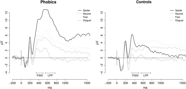
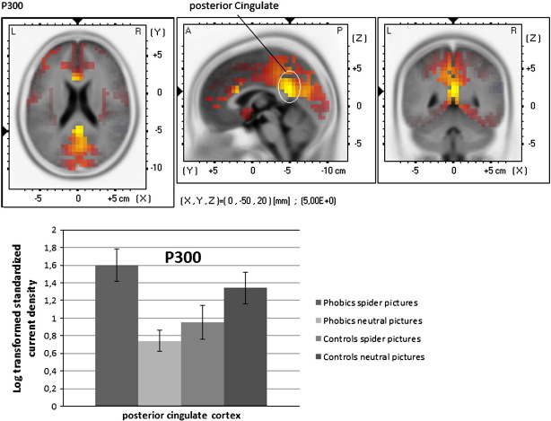
This symptom provocation study on spider phobia investigated sources of late event-related potentials (ERPs) using sLORETA (standardized low resolution brain electromagnetic tomography). Twenty-five phobic female patients and 20 non-phobic controls were confronted with phobia-relevant, generally fear-inducing, disgust-inducing and affectively neutral pictures while an electroencephalogram was recorded. Mean amplitudes of ERPs were extracted in the time windows 340-500 ms (P300) and 550-770 ms (late positive potential, LPP). Phobics showed enhanced P300 and LPP amplitudes in response to spider pictures relative to controls. Sources were mainly located in areas engaged in visuo-attentional processing (occipital and parietal regions, ventral visual pathway). Moreover, there were sources in areas which are crucial for emotional processing and the representations of aversive bodily states (cingulate cortex, insula). Further sources were located in premotor areas reflecting the priming of flight behaviour. Our findings are in good accordance with existing brain imaging studies and underline that source localization is a useful alternative for identifying phobia-relevant cortical regions.
这项蜘蛛恐惧症的症状激发研究使用 sLORETA(标准化低分辨率脑电磁层析成像)来探究晚期事件相关电位(ERP)的来源。25 名女性恐惧症患者和 20 名非恐惧症对照者在接受与恐惧症相关的、通常会引起恐惧、引起厌恶和情感中性的图片时,记录了脑电图。在 340-500 毫秒(P300)和 550-770 毫秒(晚期正电位,LPP)的时间窗口中提取 ERP 的平均幅度。与对照组相比,恐惧症患者对蜘蛛图片的反应表现出增强的 P300 和 LPP 幅度。源主要位于参与视注意处理的区域(枕叶和顶叶区域,腹侧视觉通路)。此外,还有一些位于对情绪处理和厌恶身体状态表示至关重要的区域的源(扣带皮层、脑岛)。进一步的源位于反映逃避行为启动的运动前区域。我们的发现与现有的脑成像研究一致,强调了源定位是识别恐惧症相关皮质区域的有用替代方法。