Department of Diagnostic and Interventional Radiology, Aichi Cancer Center Hospital, Nagoya 464-8681, Japan.
Korean J Radiol. 2011 May-Jun;12(3):351-7. doi: 10.3348/kjr.2011.12.3.351. Epub 2011 Apr 26.
The purpose of this study was to investigate retrospectively the clinical procedural performance of CT-guided needle biopsy for retroperitoneal lesions.
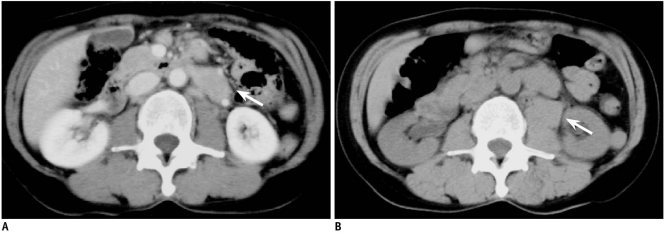
CT-guided needle biopsy was performed in 74 consecutive patients (M:F = 44:30; mean age, 59.7 years) with retroperitoneal lesions between April 1998 and June 2009. The target lesion ranged from 1.5 to 12.5 cm in size. The biopsy access path ranged from 3.5 to 11.5 cm in depth. A biopsy specimen was obtained using an 18-gauge core needle under a CT or CT-fluoroscopy guidance and with the patient under local anesthesia. The histopathological diagnoses from the biopsies were obtained. The diagnostic confirmation of the subtype of lymphoma was evaluated.
Satisfactory biopsy samples were obtained in 73 (99%) of 74 patients and a pathological diagnosis was made in 70 (95%) of 74 patients. Sixty three lesions were malignant (45 lymphomas, nine primary tumors, nine lymph node metastases) and seven were benign. The subtype of lymphoma was specified in 43 (96%) of 45 patients who were diagnosed with lymphoma. Analysis of the value of CT-guided biopsy in this series indicated 63 true positives, zero false positive, six true negatives and five false negatives. This test had a sensitivity of 93%, a specificity of 100% and an accuracy of 93%. No major complications were seen and minor complications were noted in seven patients (five with local hematomas, two with transient pain at the puncture site).
CT-guided needle biopsy for retroperitoneal lesions is highly practical and useful, and particularly for determining the subtypes in patients with lymphoma.
本研究旨在回顾性分析 CT 引导下经皮穿刺活检腹膜后病变的临床操作过程。
1998 年 4 月至 2009 年 6 月,对 74 例腹膜后病变患者(男 44 例,女 30 例;平均年龄 59.7 岁)行 CT 引导下经皮穿刺活检。目标病变大小 1.512.5cm,穿刺路径深度 3.511.5cm。在 CT 或 CT 透视引导下,局部麻醉下使用 18G 活检针获取活检标本。获得活检组织的组织病理学诊断。评估对淋巴瘤亚型的诊断确认。
74 例患者中,73 例(99%)获得满意的活检标本,74 例中有 70 例(95%)获得病理诊断。63 例病变为恶性(45 例淋巴瘤,9 例原发性肿瘤,9 例淋巴结转移),7 例为良性。45 例诊断为淋巴瘤的患者中,明确了淋巴瘤的亚型。对本系列研究中 CT 引导下活检的价值分析表明,63 例为真阳性,0 例为假阳性,6 例为真阴性,5 例为假阴性。该检查的敏感性为 93%,特异性为 100%,准确性为 93%。无重大并发症,7 例患者出现轻微并发症(5 例局部血肿,2 例穿刺部位短暂疼痛)。
CT 引导下经皮穿刺活检腹膜后病变非常实用且有效,尤其有助于确定淋巴瘤患者的亚型。