• 文献检索
  • 文档翻译
  • 深度研究
  • 学术资讯
  • Suppr Zotero 插件Zotero 插件
  • 邀请有礼
  • 套餐&价格
  • 历史记录
应用&插件
Suppr Zotero 插件Zotero 插件浏览器插件Mac 客户端Windows 客户端微信小程序
定价
高级版会员购买积分包购买API积分包
服务
文献检索文档翻译深度研究API 文档MCP 服务
关于我们
关于 Suppr公司介绍联系我们用户协议隐私条款
关注我们

Suppr 超能文献

核心技术专利:CN118964589B侵权必究
粤ICP备2023148730 号-1Suppr @ 2026

文献检索

告别复杂PubMed语法,用中文像聊天一样搜索,搜遍4000万医学文献。AI智能推荐,让科研检索更轻松。

立即免费搜索

文件翻译

保留排版,准确专业,支持PDF/Word/PPT等文件格式,支持 12+语言互译。

免费翻译文档

深度研究

AI帮你快速写综述,25分钟生成高质量综述,智能提取关键信息,辅助科研写作。

立即免费体验

Stratus光学相干断层扫描(OCT)与Spectralis OCT对视网膜神经纤维层厚度的比较。

Comparison of retinal nerve fiber layer thickness between Stratus and Spectralis OCT.

作者信息

Shin Hyun Jin, Cho Byung Joo

机构信息

Department of Ophthalmology, Konkuk University School of Medicine, Seoul, Korea.

出版信息

Korean J Ophthalmol. 2011 Jun;25(3):166-73. doi: 10.3341/kjo.2011.25.3.166. Epub 2011 May 24.

DOI:10.3341/kjo.2011.25.3.166
PMID:21655041
原文链接:https://pmc.ncbi.nlm.nih.gov/articles/PMC3102819/
Abstract

PURPOSE

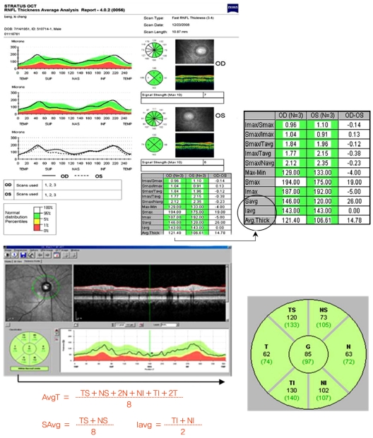
To compare the peripapillary retinal nerve fiber layer (RNFL) thickness of normal patients and those with various glaucoma diseases by time domain (Stratus) and spectral domain (Spectralis) optical coherence tomography (OCT).

METHODS

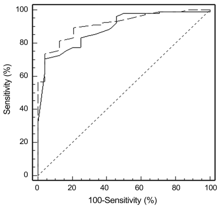
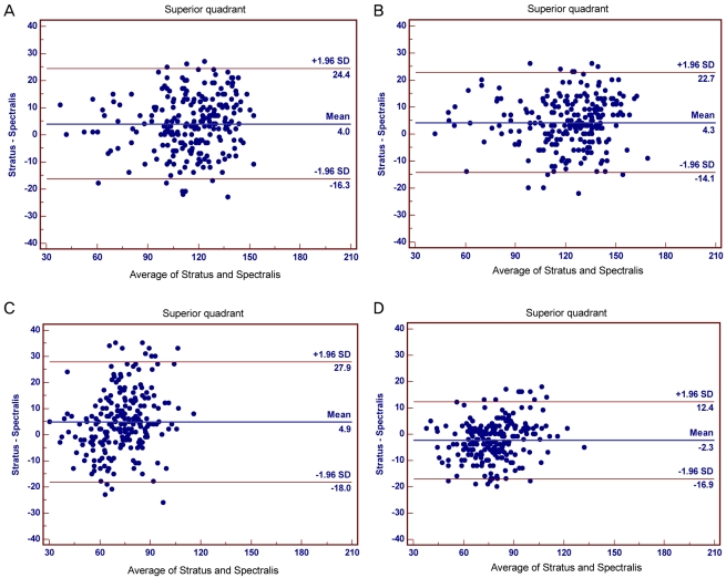
The RNFL thickness as measured by the Stratus and Spectral OCT was compared (paired t-test). The relationship and agreement of RNFL thickness between the two OCT modalities were evaluated by Pearson correlation, Bland-Altman plot, and area under the receiver operating characteristic curve.

RESULTS

Two-hundred seventeen eyes of 217 patients, including twenty-four normal eyes, ninety-one glaucoma suspects, seventy-six normal tension glaucoma cases, and twenty-six primary open angle glaucoma cases (POAG) were analyzed. The peripapillary RNFL thicknesses as measured by Stratus OCT were significantly greater than those measured by Spectralis OCT. However, in quadrant comparisons, the temporal RNFL thickness obtained using Stratus OCT were significantly less than those obtained using Spectralis OCT. Correlations between RNFL parameters were strong (Pearson correlation coefficient for mean RNFL thickness = 0.88); a high degree of correlation was found in the POAG group. Bland-Altman plotting demonstrated that agreement in the temporal quadrant was greater than any other quadrant.

CONCLUSIONS

Both OCT systems were highly correlated and demonstrated strong agreement. However, absolute measurements of peripapillary RNFL thickness differed between Stratus OCT and Spectralis OCT. Thus, measurements with these instruments should not be considered interchangeable. The temporal quadrant was the only sector where RNFL thickness as measured by Spectralis OCT was greater than by Stratus OCT; this demonstrated greater agreement than other sectors.

摘要

目的

通过时域(Stratus)和频域(Spectralis)光学相干断层扫描(OCT)比较正常患者与患有各种青光眼疾病患者的视乳头周围视网膜神经纤维层(RNFL)厚度。

方法

比较Stratus和Spectral OCT测量的RNFL厚度(配对t检验)。通过Pearson相关性、Bland-Altman图和受试者操作特征曲线下面积评估两种OCT模式之间RNFL厚度的关系和一致性。

结果

分析了217例患者的217只眼,包括24只正常眼、91只青光眼疑似病例、76只正常眼压性青光眼病例和26只原发性开角型青光眼病例(POAG)。Stratus OCT测量的视乳头周围RNFL厚度显著大于Spectralis OCT测量的厚度。然而,在象限比较中,Stratus OCT获得的颞侧RNFL厚度显著小于Spectralis OCT获得的厚度。RNFL参数之间的相关性很强(平均RNFL厚度的Pearson相关系数 = 0.88);在POAG组中发现高度相关性。Bland-Altman图显示颞侧象限的一致性大于其他任何象限。

结论

两种OCT系统高度相关且显示出很强的一致性。然而,Stratus OCT和Spectralis OCT对视乳头周围RNFL厚度的绝对测量值不同。因此,不应认为使用这些仪器进行的测量是可互换的。颞侧象限是Spectralis OCT测量的RNFL厚度大于Stratus OCT测量厚度的唯一象限;这表明该象限的一致性高于其他象限。

https://cdn.ncbi.nlm.nih.gov/pmc/blobs/f8d2/3102819/fc378bc35715/kjo-25-166-g003.jpg
https://cdn.ncbi.nlm.nih.gov/pmc/blobs/f8d2/3102819/71a1cefcc463/kjo-25-166-g001.jpg
https://cdn.ncbi.nlm.nih.gov/pmc/blobs/f8d2/3102819/689ab285c996/kjo-25-166-g002.jpg
https://cdn.ncbi.nlm.nih.gov/pmc/blobs/f8d2/3102819/fc378bc35715/kjo-25-166-g003.jpg
https://cdn.ncbi.nlm.nih.gov/pmc/blobs/f8d2/3102819/71a1cefcc463/kjo-25-166-g001.jpg
https://cdn.ncbi.nlm.nih.gov/pmc/blobs/f8d2/3102819/689ab285c996/kjo-25-166-g002.jpg
https://cdn.ncbi.nlm.nih.gov/pmc/blobs/f8d2/3102819/fc378bc35715/kjo-25-166-g003.jpg

相似文献

1
Comparison of retinal nerve fiber layer thickness between Stratus and Spectralis OCT.Stratus光学相干断层扫描(OCT)与Spectralis OCT对视网膜神经纤维层厚度的比较。
Korean J Ophthalmol. 2011 Jun;25(3):166-73. doi: 10.3341/kjo.2011.25.3.166. Epub 2011 May 24.
2
Comparison of the diagnostic ability of retinal nerve fiber layer thickness measured using time domain and spectral domain optical coherence tomography in primary open angle glaucoma.时域和频域光学相干断层扫描测量视网膜神经纤维层厚度在原发性开角型青光眼中的诊断能力比较
Eye Sci. 2011 Sep;26(3):132-7, 142. doi: 10.3969/j.issn.1000-4432.2011.03.002.
3
Comparison of retinal nerve fiber layer measurements using time domain and spectral domain optical coherent tomography.使用时域和频域光学相干断层扫描技术测量视网膜神经纤维层的比较。
Ophthalmology. 2009 Jul;116(7):1271-7. doi: 10.1016/j.ophtha.2008.12.032. Epub 2009 Apr 22.
4
Retinal nerve fiber layer measurement and diagnostic capability of spectral-domain versus time-domain optical coherence tomography.光谱域与时域光学相干断层扫描对视网膜神经纤维层的测量及诊断能力
Eur J Ophthalmol. 2011 Sep-Oct;21(5):566-72. doi: 10.5301/EJO.2011.6289.
5
Comparative evaluation of time domain and spectral domain optical coherence tomography in retinal nerve fiber layer thickness measurements.视网膜神经纤维层厚度测量中时域与频域光学相干断层扫描的比较评估
Nepal J Ophthalmol. 2014 Jul-Dec;6(2):185-91. doi: 10.3126/nepjoph.v6i2.11692.
6
Reproducibility and agreement in evaluating retinal nerve fibre layer thickness between Stratus and Spectralis OCT.Stratus 和 Spectralis OCT 评估视网膜神经纤维层厚度的可重复性和一致性。
Eye (Lond). 2011 Feb;25(2):192-200. doi: 10.1038/eye.2010.178. Epub 2010 Nov 26.
7
Cirrus high-definition optical coherence tomography compared with Stratus optical coherence tomography in glaucoma diagnosis.脉络膜高清光学相干断层扫描与 Stratus 光学相干断层扫描在青光眼诊断中的比较。
Invest Ophthalmol Vis Sci. 2010 Jan;51(1):335-43. doi: 10.1167/iovs.08-2988. Epub 2009 Sep 8.
8
Retinal nerve fiber layer imaging with spectral-domain optical coherence tomography: a variability and diagnostic performance study.光谱域光学相干断层扫描技术对视网膜神经纤维层的成像:变异性与诊断性能研究
Ophthalmology. 2009 Jul;116(7):1257-63, 1263.e1-2. doi: 10.1016/j.ophtha.2009.04.013. Epub 2009 May 22.
9
Comparison of retinal nerve fiber layer thickness values using Stratus Optical Coherence Tomography and Heidelberg Retina Tomograph-III.使用Stratus光学相干断层扫描和海德堡视网膜断层扫描仪III比较视网膜神经纤维层厚度值。
J Glaucoma. 2009 Sep;18(7):528-34. doi: 10.1097/IJG.0b013e318193c29f.
10
Comparison of ganglion cell and retinal nerve fiber layer thickness in primary open-angle glaucoma and normal tension glaucoma with spectral-domain OCT.应用谱域光学相干断层扫描比较原发性开角型青光眼和正常眼压型青光眼的神经节细胞和视网膜神经纤维层厚度。
Graefes Arch Clin Exp Ophthalmol. 2013 Mar;251(3):831-8. doi: 10.1007/s00417-012-2114-5. Epub 2012 Aug 18.

引用本文的文献

1
Anti-vascular endothelial growth factor drugs combined with laser photocoagulation maintain retinal ganglion cell integrity in patients with diabetic macular edema: study protocol for a prospective, non-randomized, controlled clinical trial.抗血管内皮生长因子药物联合激光光凝术可维持糖尿病性黄斑水肿患者视网膜神经节细胞的完整性:一项前瞻性、非随机、对照临床试验的研究方案
Neural Regen Res. 2024 Apr;19(4):923-928. doi: 10.4103/1673-5374.382104.
2
Phenomenology of spontaneous closure in degenerative and mixed type lamellar macular hole.特发性退行性和混合性板层黄斑裂孔自发闭合的现象学。
Eye (Lond). 2024 Feb;38(2):315-320. doi: 10.1038/s41433-023-02681-y. Epub 2023 Jul 31.
3

本文引用的文献

1
Retinal nerve fiber layer thickness in normals measured by spectral domain OCT.正常人的视网膜神经纤维层厚度的光谱域 OCT 测量。
J Glaucoma. 2010 Sep;19(7):475-82. doi: 10.1097/IJG.0b013e3181c4b0c7.
2
Comparison of retinal nerve fiber layer thickness measured by Cirrus HD and Stratus optical coherence tomography.Cirrus HD和Stratus光学相干断层扫描测量视网膜神经纤维层厚度的比较。
Ophthalmology. 2009 Jul;116(7):1264-70, 1270.e1. doi: 10.1016/j.ophtha.2008.12.045. Epub 2009 May 8.
3
Comparison of retinal nerve fiber layer measurements using time domain and spectral domain optical coherent tomography.
Optical Coherence Tomography: Is Really a New Biomarker for Alzheimer's Disease?
光学相干断层扫描:真的是阿尔茨海默病的一种新生物标志物吗?
Ann Indian Acad Neurol. 2018 Apr-Jun;21(2):119-125. doi: 10.4103/aian.AIAN_368_17.
4
Investigation of lateral geniculate nucleus volume and diffusion tensor imaging in patients with normal tension glaucoma using 7 tesla magnetic resonance imaging.使用 7 特斯拉磁共振成像研究正常眼压性青光眼患者外侧膝状体核体积和弥散张量成像。
PLoS One. 2018 Jun 7;13(6):e0198830. doi: 10.1371/journal.pone.0198830. eCollection 2018.
5
Optical coherence tomography for glaucoma diagnosis: An evidence based meta-analysis.用于青光眼诊断的光学相干断层扫描:一项基于证据的荟萃分析。
PLoS One. 2018 Jan 4;13(1):e0190621. doi: 10.1371/journal.pone.0190621. eCollection 2018.
6
Three-dimensional mapping of peripapillary retinal layers using a spectral domain optical coherence tomography.使用光谱域光学相干断层扫描对视乳头周围视网膜层进行三维映射。
Clin Ophthalmol. 2017 Dec 11;11:2191-2198. doi: 10.2147/OPTH.S150617. eCollection 2017.
7
Retinal Nerve Fiber Layer Thickness Measurement Comparison Using Spectral Domain and Swept Source Optical Coherence Tomography.使用光谱域和扫频光学相干断层扫描技术比较视网膜神经纤维层厚度测量
Korean J Ophthalmol. 2016 Apr;30(2):140-7. doi: 10.3341/kjo.2016.30.2.140. Epub 2016 Mar 25.
8
Comparison of retinal nerve fiber layer and macular thickness measurements with Stratus OCT and OPKO/OTI OCT devices in healthy subjects.健康受试者中使用Stratus OCT和OPKO/OTI OCT设备测量视网膜神经纤维层和黄斑厚度的比较。
Int J Ophthalmol. 2015 Feb 18;8(1):98-103. doi: 10.3980/j.issn.2222-3959.2015.01.18. eCollection 2015.
9
Effects of multiple intravitreal anti-VEGF injections on retinal nerve fiber layer and intraocular pressure: a comparative clinical study.多次玻璃体内注射抗血管内皮生长因子对视网膜神经纤维层和眼压的影响:一项对比临床研究
Int J Ophthalmol. 2014 Aug 18;7(4):740. doi: 10.3980/j.issn.2222-3959.2014.04.30. eCollection 2014.
10
Analysis of peripapilary retinal nerve fiber layer thickness of healthy Chinese from northwestern Shanghai using Cirrus HD-OCT.使用Cirrus HD-OCT对上海西北部健康中国人的视乳头周围视网膜神经纤维层厚度进行分析。
Int J Ophthalmol. 2014 Aug 18;7(4):654-8. doi: 10.3980/j.issn.2222-3959.2014.04.12. eCollection 2014.
使用时域和频域光学相干断层扫描技术测量视网膜神经纤维层的比较。
Ophthalmology. 2009 Jul;116(7):1271-7. doi: 10.1016/j.ophtha.2008.12.032. Epub 2009 Apr 22.
4
Comparison of spectral- and time-domain optical coherence tomography for retinal thickness measurements in healthy and diseased eyes.光谱域和时域光学相干断层扫描技术在健康和患病眼睛视网膜厚度测量中的比较。
Am J Ophthalmol. 2009 May;147(5):847-58, 858.e1. doi: 10.1016/j.ajo.2008.11.019. Epub 2009 Feb 6.
5
Relationship between retinal nerve fiber layer measurement and signal strength in optical coherence tomography.光学相干断层扫描中视网膜神经纤维层测量与信号强度之间的关系。
Ophthalmology. 2008 Aug;115(8):1347-51, 1351.e1-2. doi: 10.1016/j.ophtha.2007.11.027. Epub 2008 Feb 21.
6
Retinal nerve fibre layer imaging compared with histological measurements in a human eye.人眼视网膜神经纤维层成像与组织学测量结果的比较
Eye (Lond). 2009 Jan;23(1):171-5. doi: 10.1038/sj.eye.6702942. Epub 2007 Aug 24.
7
Optical coherence tomography and histologic measurements of nerve fiber layer thickness in normal and glaucomatous monkey eyes.正常和青光眼猴眼神经纤维层厚度的光学相干断层扫描及组织学测量
Invest Ophthalmol Vis Sci. 2007 Aug;48(8):3645-54. doi: 10.1167/iovs.06-0876.
8
Factors associated with variability in retinal nerve fiber layer thickness measurements obtained by optical coherence tomography.光学相干断层扫描获得的视网膜神经纤维层厚度测量值变异性的相关因素。
Ophthalmology. 2007 Aug;114(8):1505-12. doi: 10.1016/j.ophtha.2006.10.061. Epub 2007 Mar 23.
9
Ultra-high speed and ultra-high resolution spectral-domain optical coherence tomography and optical Doppler tomography in ophthalmology.眼科中的超高速和超高分辨率光谱域光学相干断层扫描及光学多普勒断层扫描
Bull Soc Belge Ophtalmol. 2006(302):123-32.
10
Spectral domain optical coherence tomography: ultra-high speed, ultra-high resolution ophthalmic imaging.频域光学相干断层扫描:超高速、超高分辨率眼科成像。
Arch Ophthalmol. 2005 Dec;123(12):1715-20. doi: 10.1001/archopht.123.12.1715.